Self-learning and aPKC
on Monday, July 13th, 2026 8:37 | by Björn Brembs
In this paper on Aplysia operant conditioning, Fred Lorenzetti and colleagues find that PKC activity is upstream of an Adenylyl cyklase making cAMP (with other hypotheses dimmed). In Drosophila, we have a different PKC (aPKC) than in Aplysia, but this may be due to aPKC having evolved after the split (needs to be tested). Dopamine, in Drosophila may be used for regulation, not convergence. This would entail some hypotheses worth testing.

According to the RNAseq database, the adenylyl cyklase rutabaga is also expressed in the motor neurons where we think the plasticity for operant self-learning takes place. We know that mutations in the rut gene improve self-learning, excluding the hypotheiss that rut may be downstream of aPKC. However, Lorenzetti et al. found that their Aplysia AC in question was a type II AC, while rut is a type I AC. It is conceivable that removal of rut could free its ATP pool for the type II AC and improve learning.
At the same time, it may be that this type II AC is downstream of a D2 receptor receiving input from the MBs inhibiting this AC, leading to inhibited self-learning while predictive colors are present. Imaging cAMP in these neurons would help us test this hypothesis.
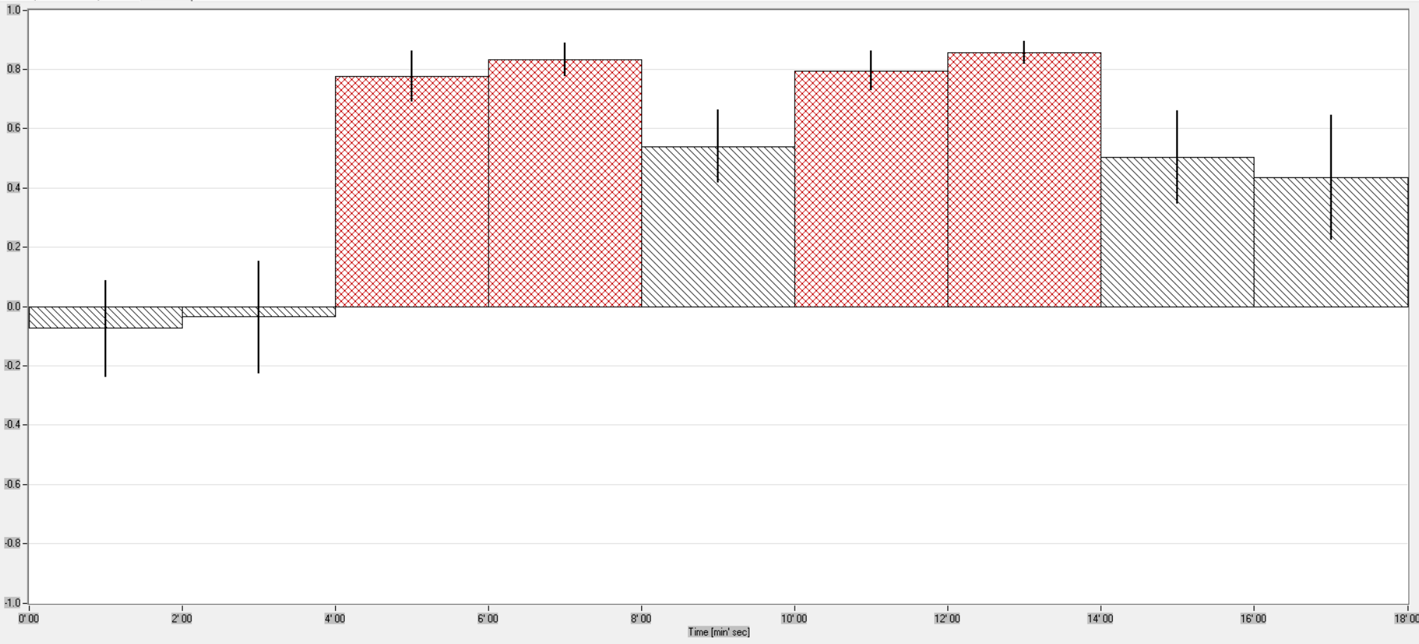
Alternatively, hypothetically, aPKC may be downstream of an inhibitory cAMP pathway, perhaps via rut, such that rut mutation would lead to improved self-learning and rut activation via D1 receptors would inhibit self-learning. This is not impossible, as rut mutant flies show premature habit formation (N=30):

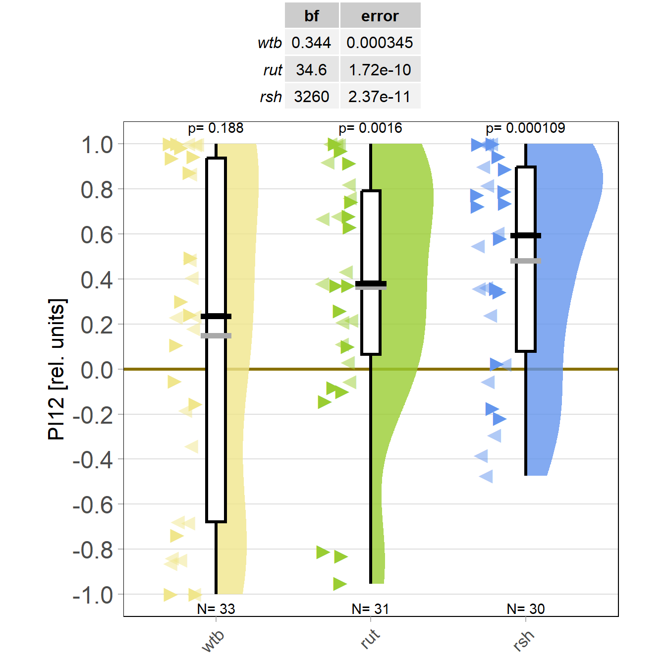
In fact, radish shows the same phenotype (N=13):
So we need a hypothesis that explains these findings:
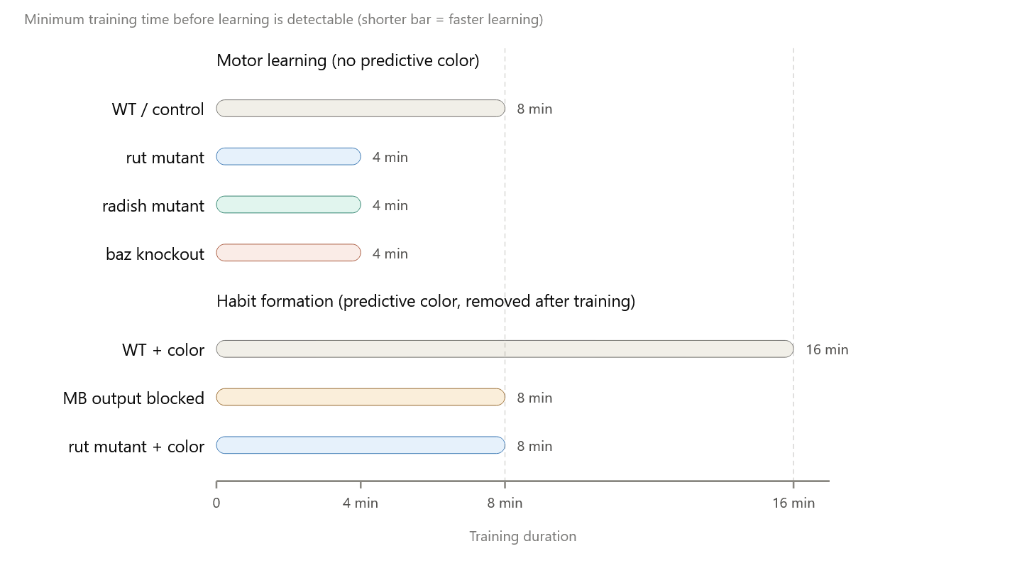
The rutabaga and radish genes inhibit self-learning (only 4 minutes training in mutants or 8 minutes with colors)
The mushroom-bodies inhibit self-learning when predictive colors are present (16 minutes training with colors)
In general, this is the problem we are trying to solve:
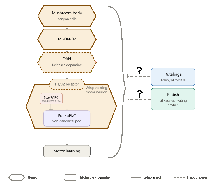
One way to test this is to do double manipulations of any of these individual ones. Another could be to quantify aPKC activity in some way. Overexpress dunce in FoxP neurons? Or wait for Julias project to give us some clues. What else?
Overview of results (maybe not complete?):

Category: Habit formation, MBON, operant self-learning, PKC | No Comments
Summary lab work
on Monday, June 22nd, 2026 12:12 | by Julia Schulz
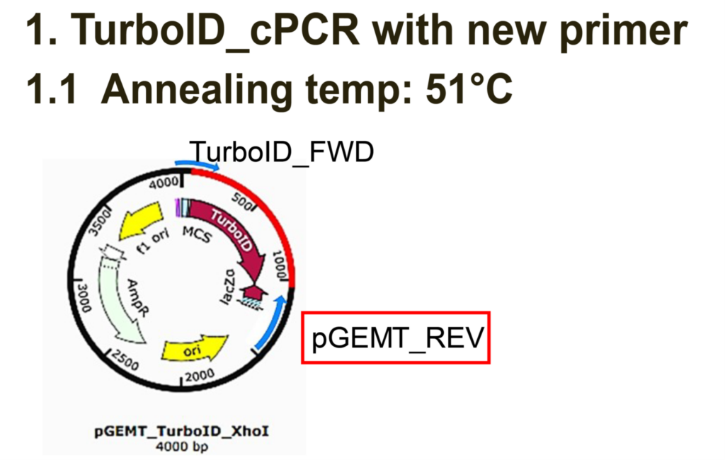
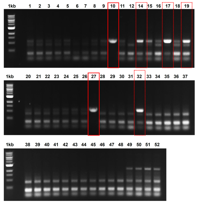
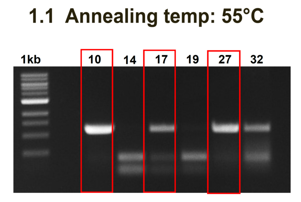
Category: PKC, TurboID-aPKC construct generation | No Comments
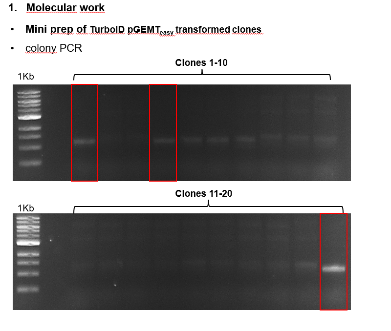
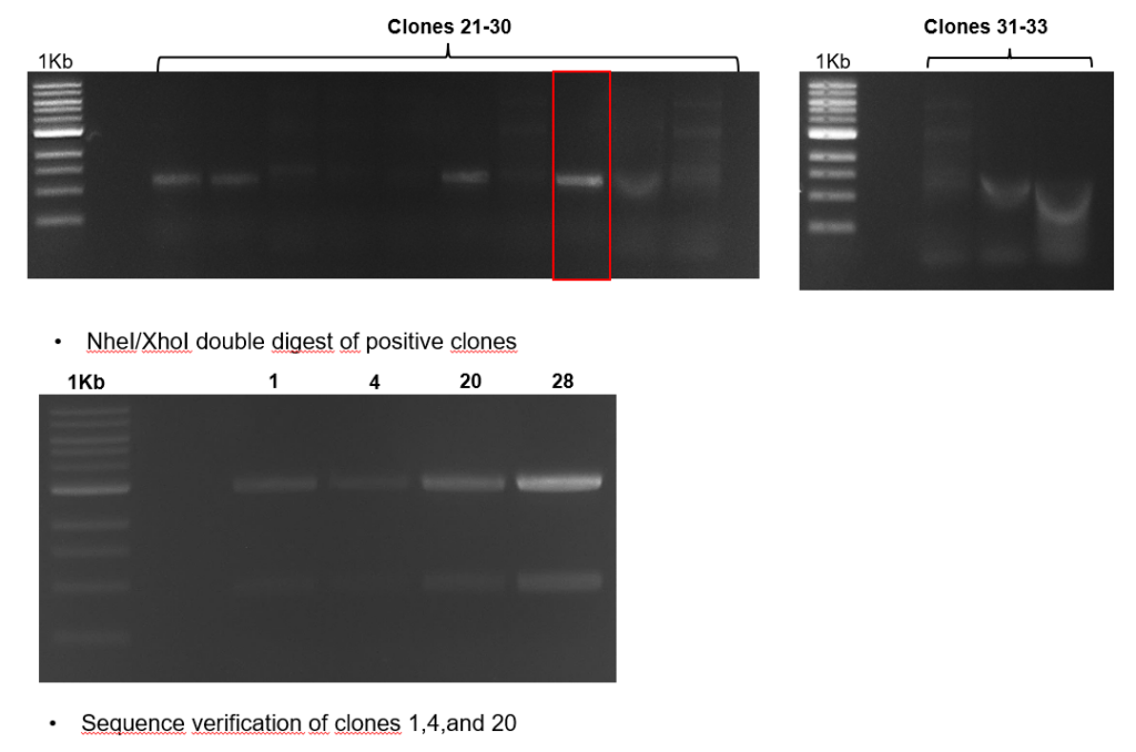
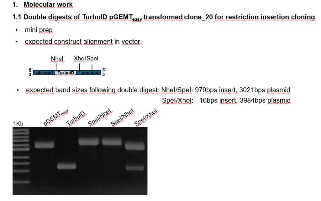
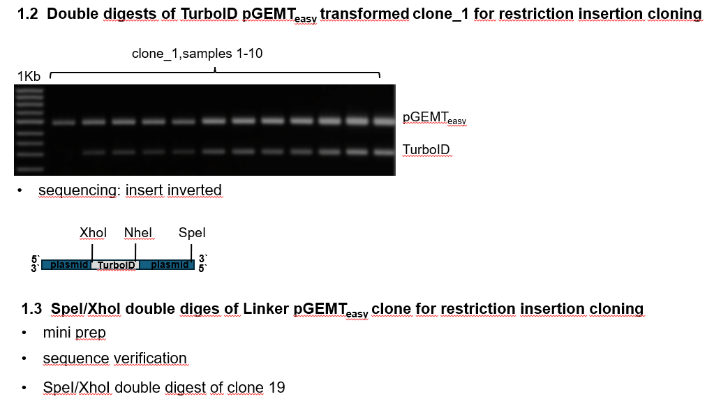
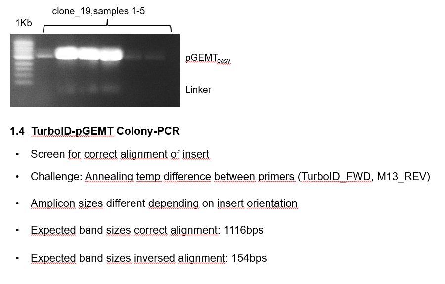
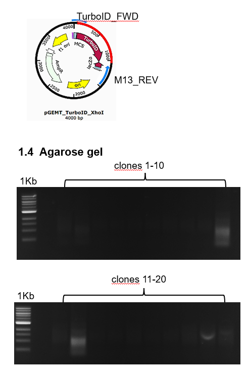
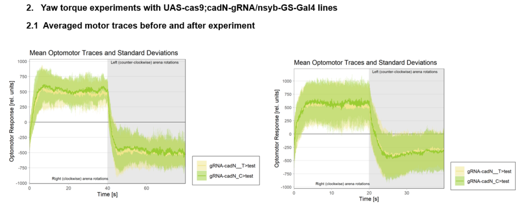
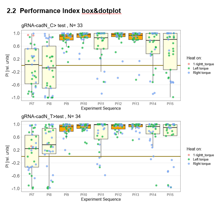
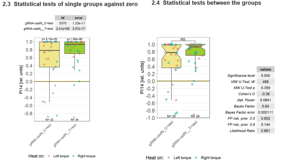
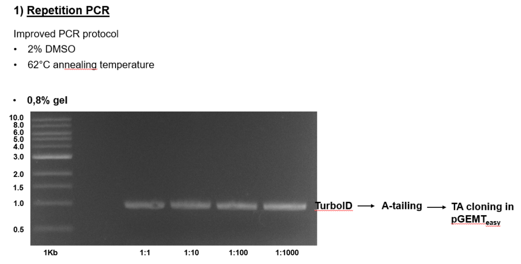
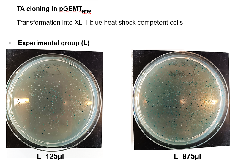
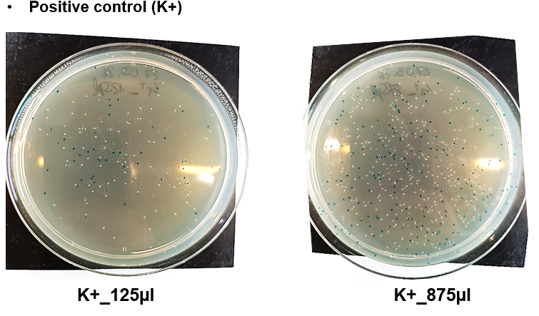
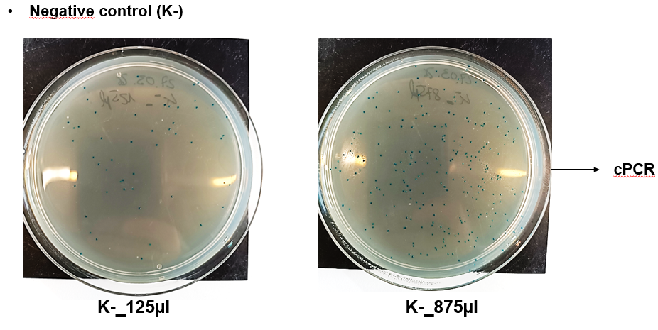
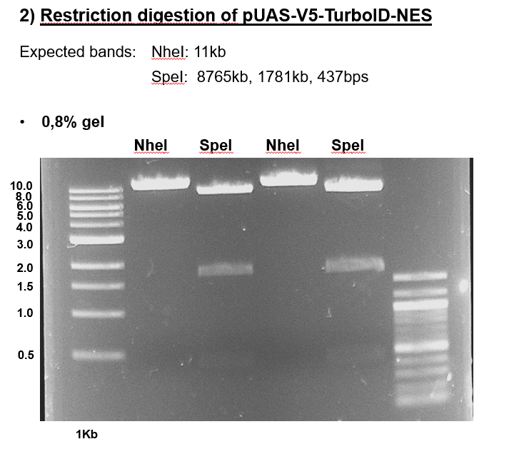
Summary molecular work
on Monday, March 30th, 2026 12:54 | by Julia Schulz
Category: Operant learning, PKC, TurboID-aPKC construct generation | No Comments
aPKC/FoxP colocalization confocal images
on Friday, January 23rd, 2026 12:52 | by Fridrik Kjartansson
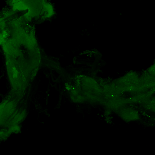
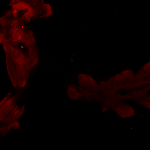
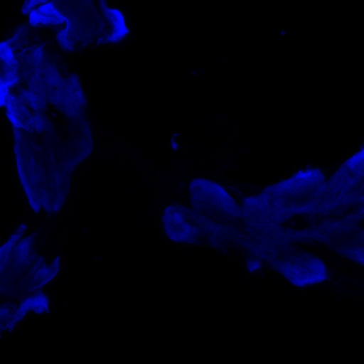
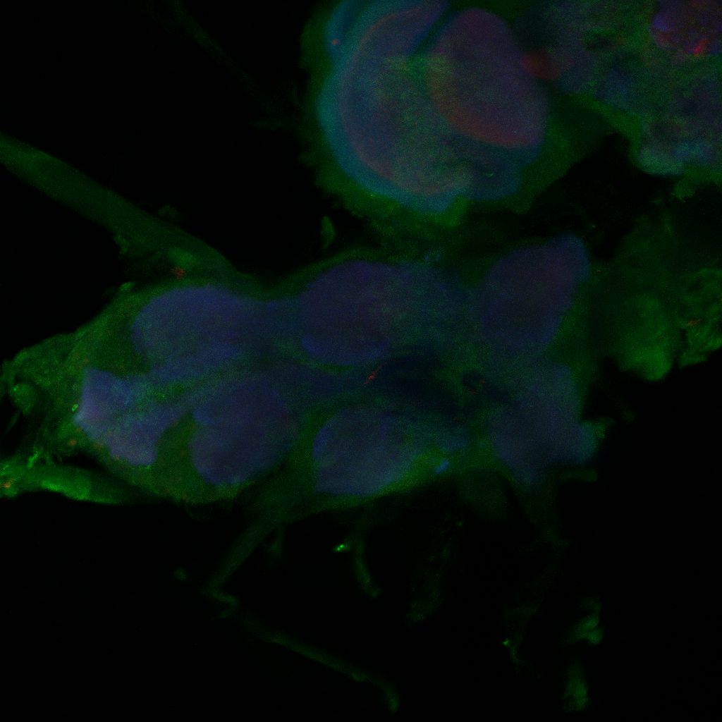
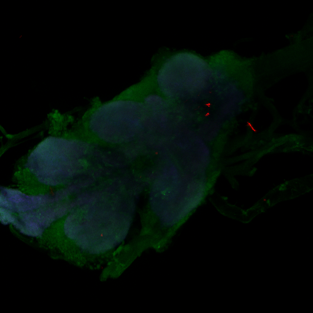
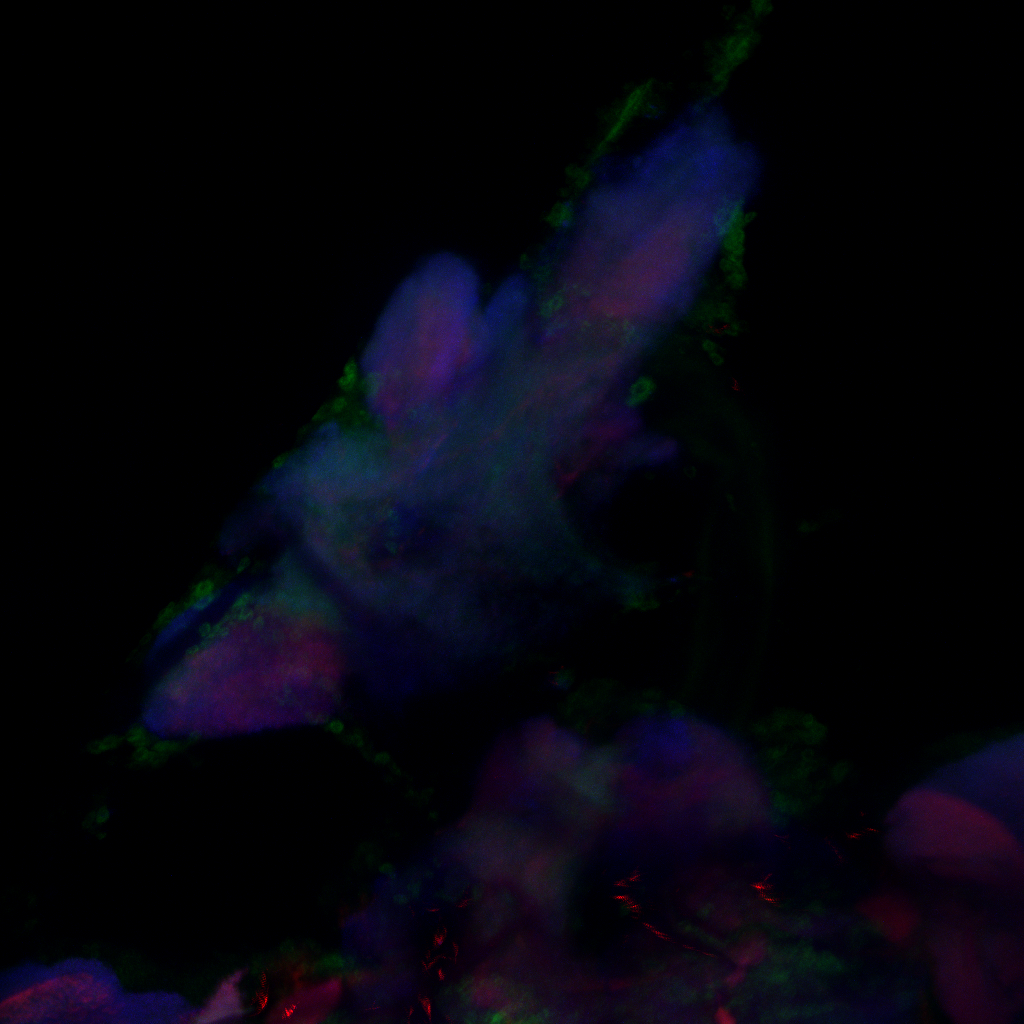
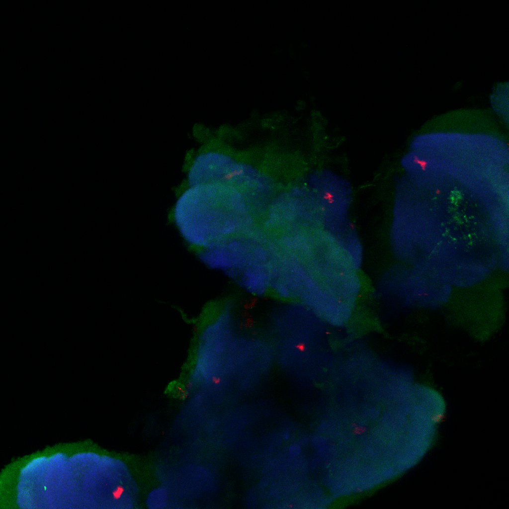
Staining is a bit faint and brain quality a bit sub-optimal and no punctae can be observed in the FoxP channel, decreasing the gain did not resolve this, blocking was done for 1 hour at RT. Perhaps increasing concentration of 1st degree antibodies, especially for RFP might improve the results.
Category: Anatomy, crosses, Foxp, PKC, PKC_localisation | No Comments
Confocal images, gain reduced
on Monday, December 1st, 2025 12:42 | by Fridrik Kjartansson
Category: Anatomy, Foxp, genetics, Operant learning, PKC, PKC_localisation | No Comments
Work flow molecular study
on Monday, November 3rd, 2025 10:57 | by Julia Schulz


Category: Operant learning, PKC | No Comments
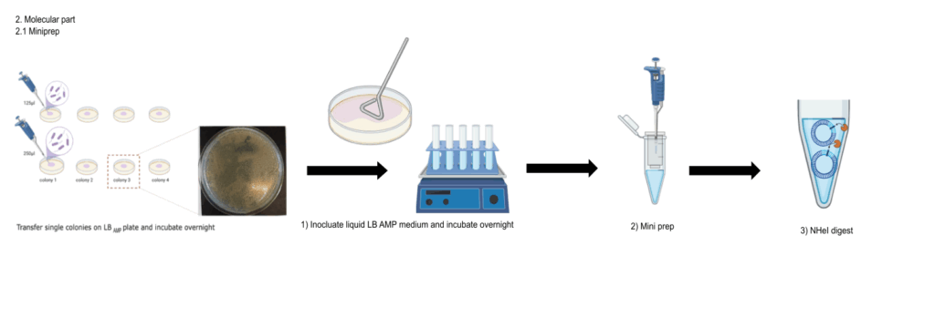
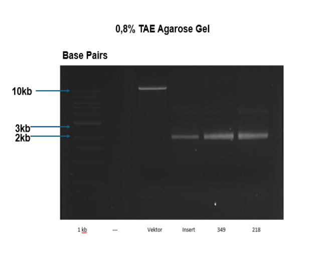
Molecular work
on Monday, August 4th, 2025 1:59 | by Julia Schulz
Category: Operant learning, PKC | No Comments
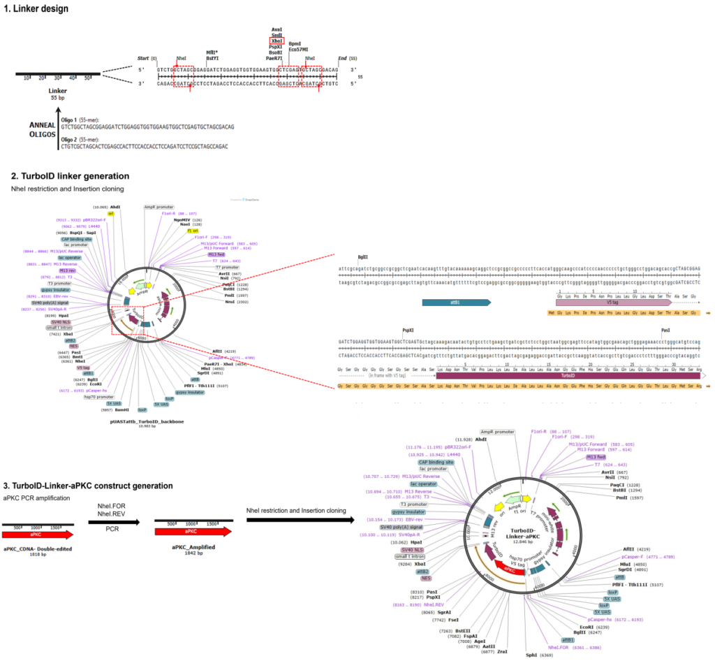
Generation of UAS-aPKC-TurboID lines
on Monday, March 24th, 2025 1:44 | by Julia Schulz
Category: operant self-learning, PKC | No Comments
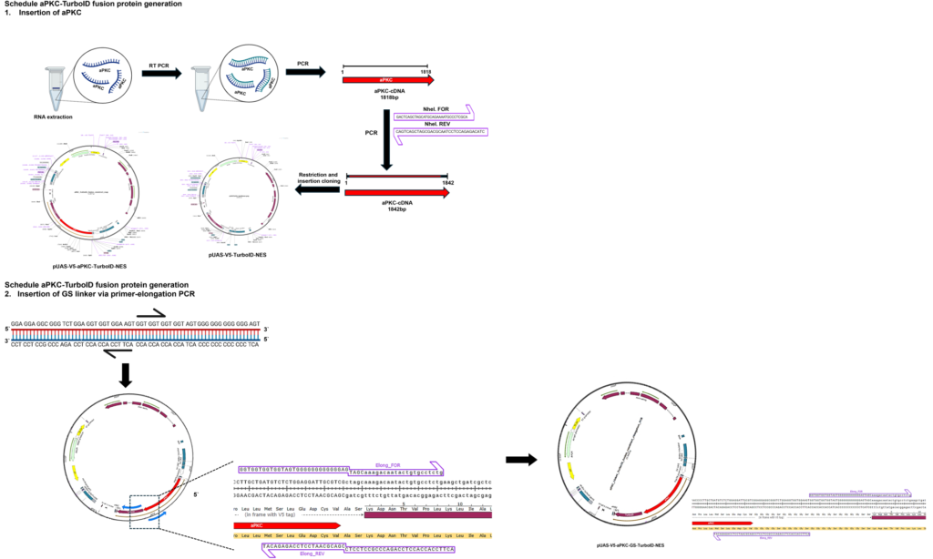
Generation of UAS-aPKC-turboID lines overview
on Monday, March 17th, 2025 1:14 | by Julia Schulz
Category: operant self-learning, PKC | No Comments