on Monday, September 11th, 2023 9:52 |
by Luisa Guyton
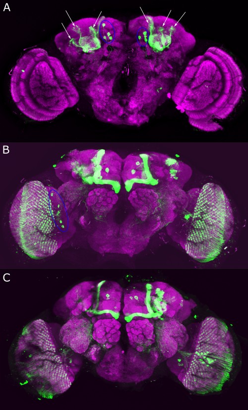
Confocal images with anti-GFP and anti-Brp staining of TH-C-AD;TH-D-DBD > mCD::GFP fly brains: A: 4 PPM 2 DANs per hemisphere marked with blue circles, three clusters of Kenyon cell bodies per hemisphere marked with white arrows. B: Five visible cell bodies of unidentified neurons marked with a blue circle in the left hemisphere. The neurons project into the lobula plate and the medulla. Strong fluorescence of cell bodies of the Kenyon cells projecting into the Mushroom bodies is visible. C: Unidentified neurons projecting into structures outside of the optic lobes especially the ventral lateral protocerebrum.

Leave a Reply