Stainning c105;;c232
on Monday, May 14th, 2018 11:22 | by Christian Rohrsen
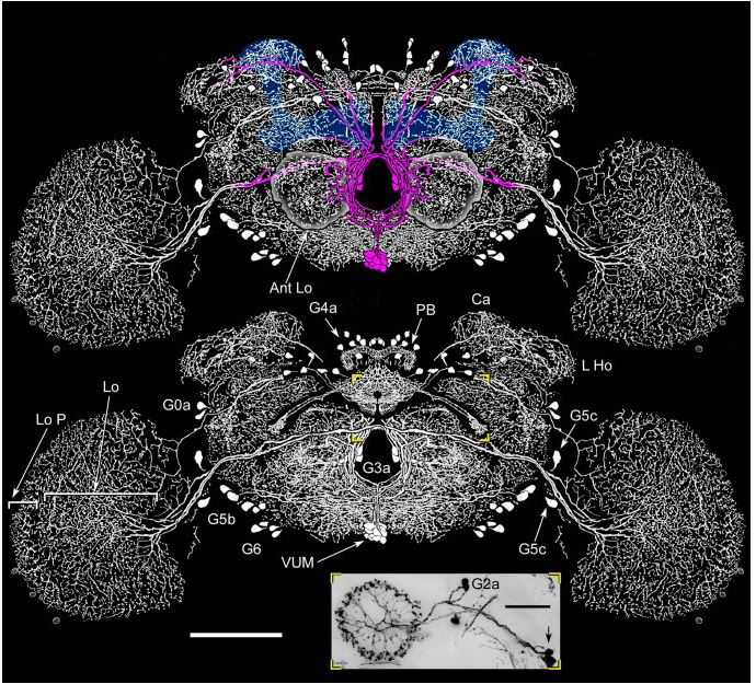
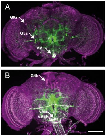
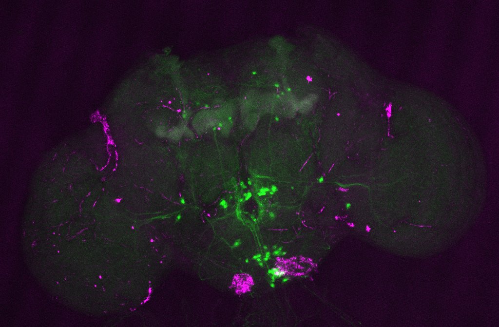
The first figure shows each of the central complex ring neurons types (Martín Pena et al., 2014). The c105-G4 targets the R1 neurons and the c232-G4 targets the R2 and the R4d neurons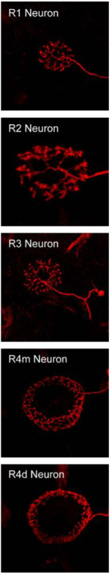
This is the c105-G4 stainning from Martín Pena et al., 2014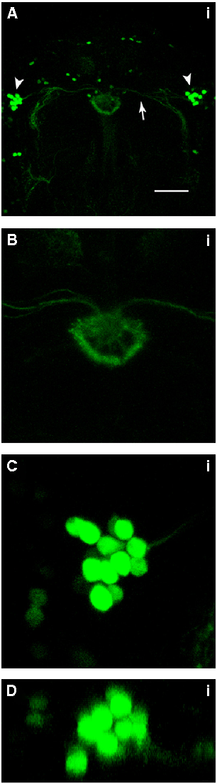
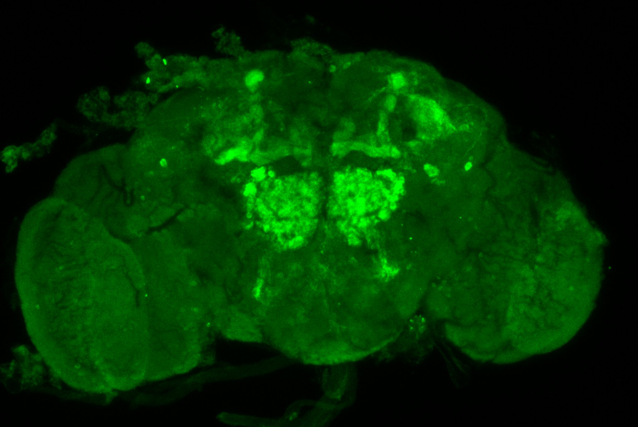
232-G4 stainning from Kahsai et al., 2012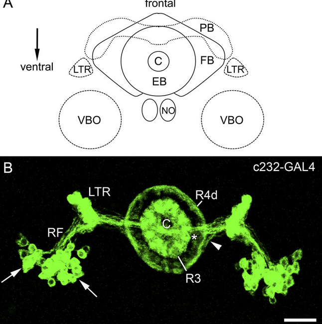
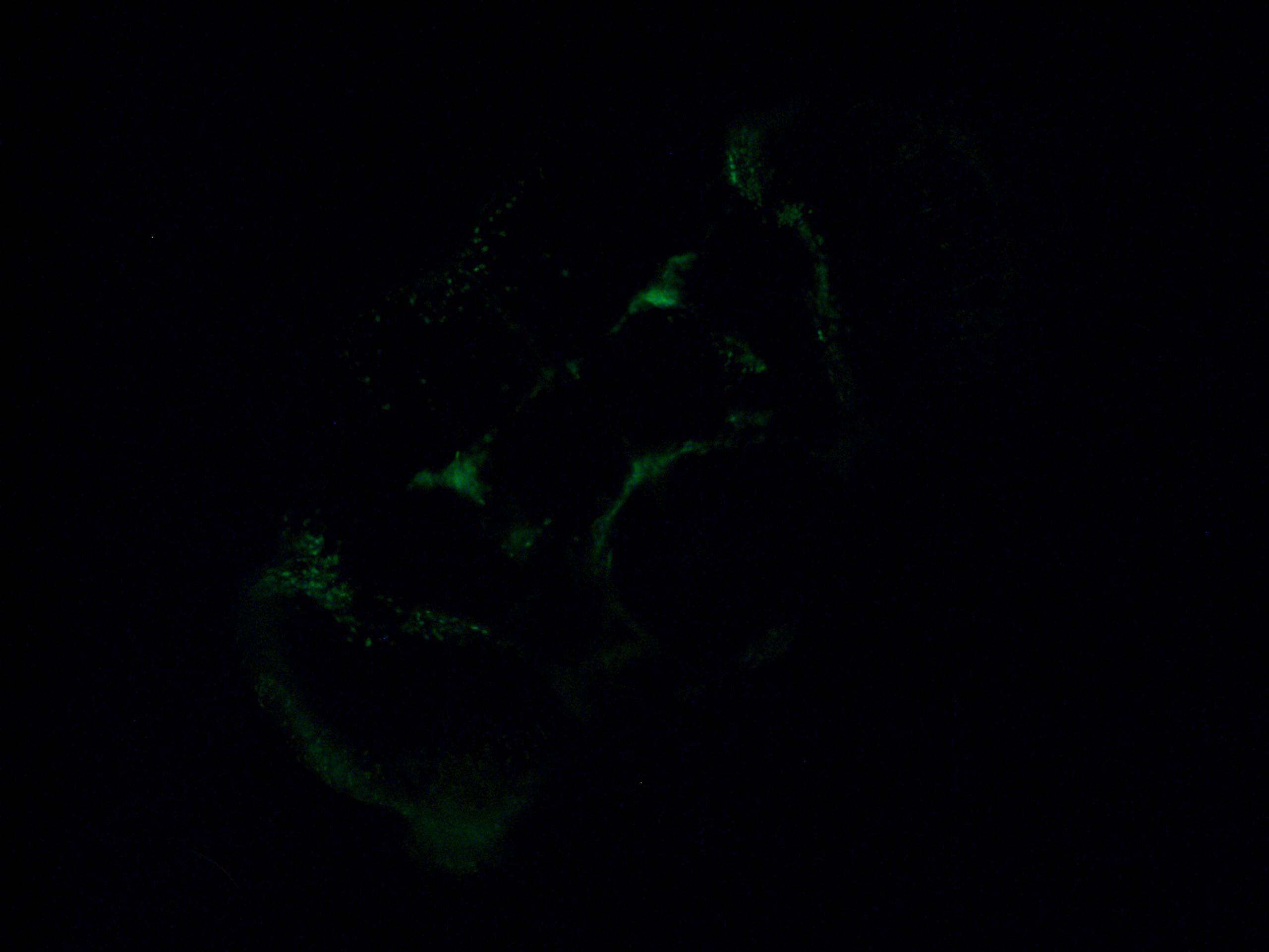
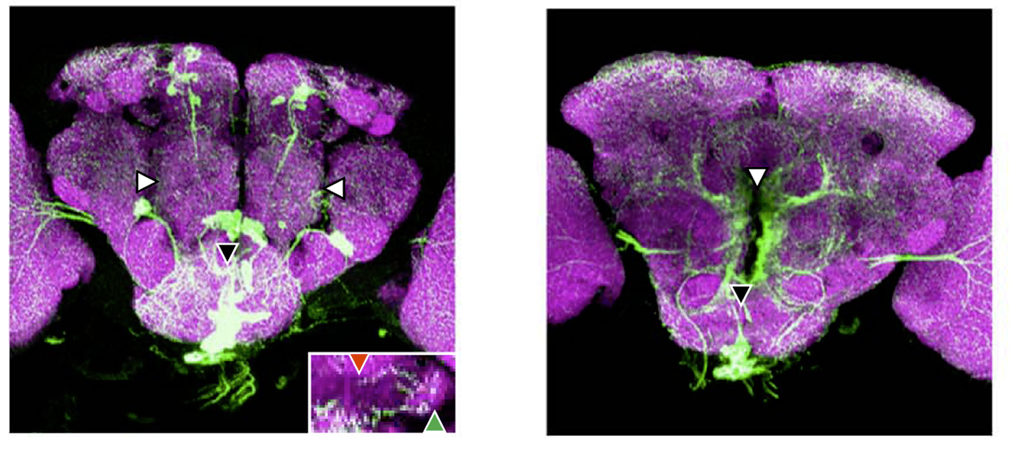
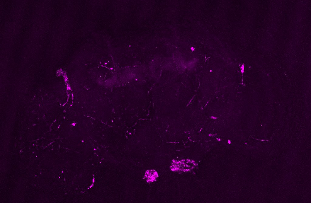
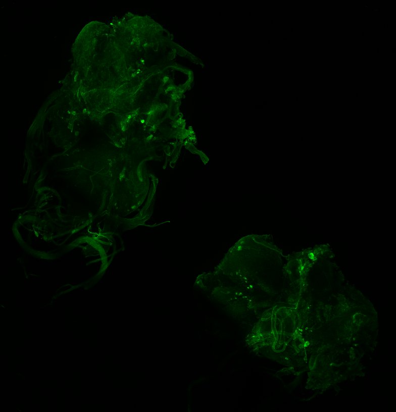
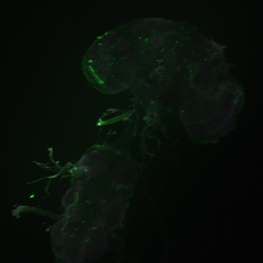
Axel stainning from c232-G4 alone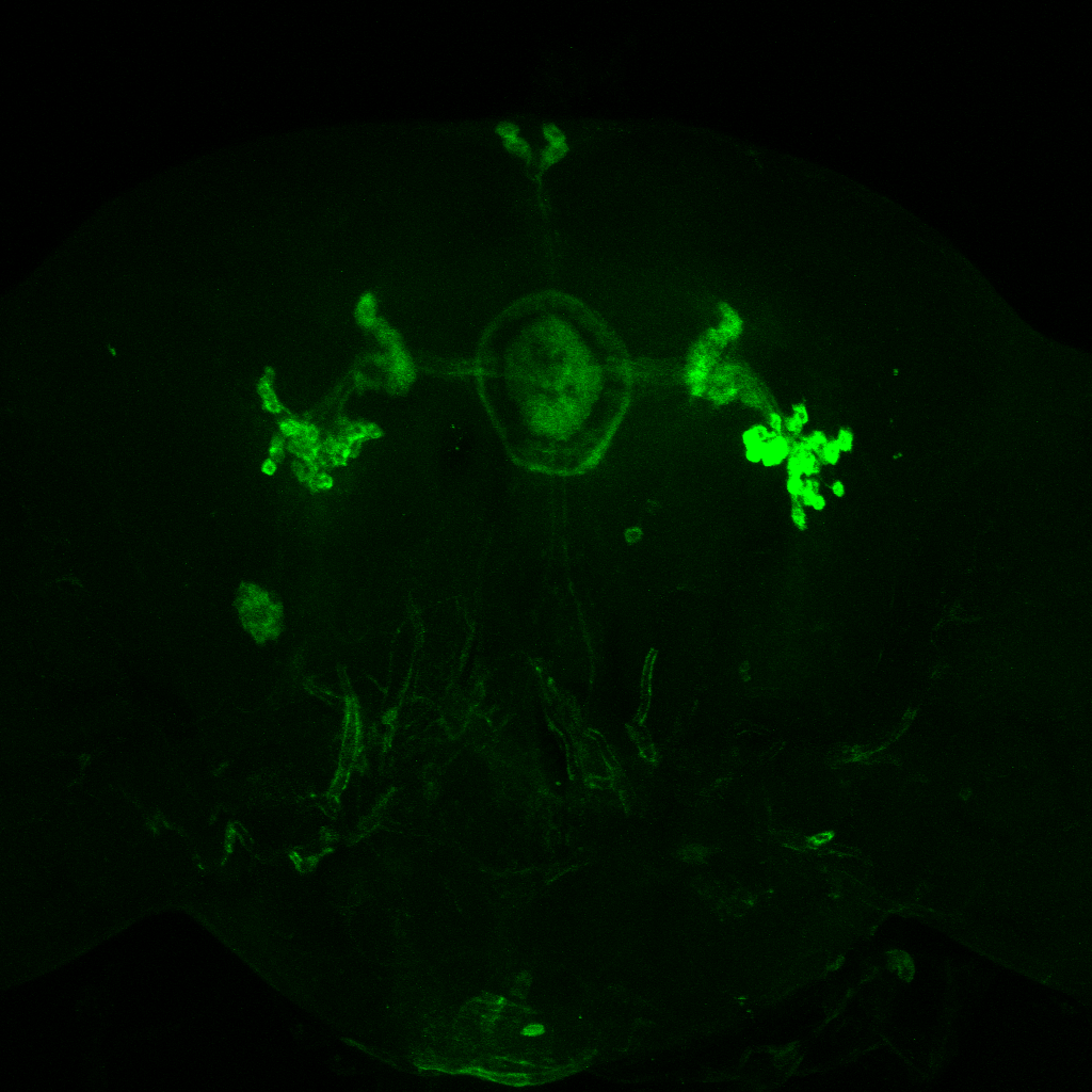
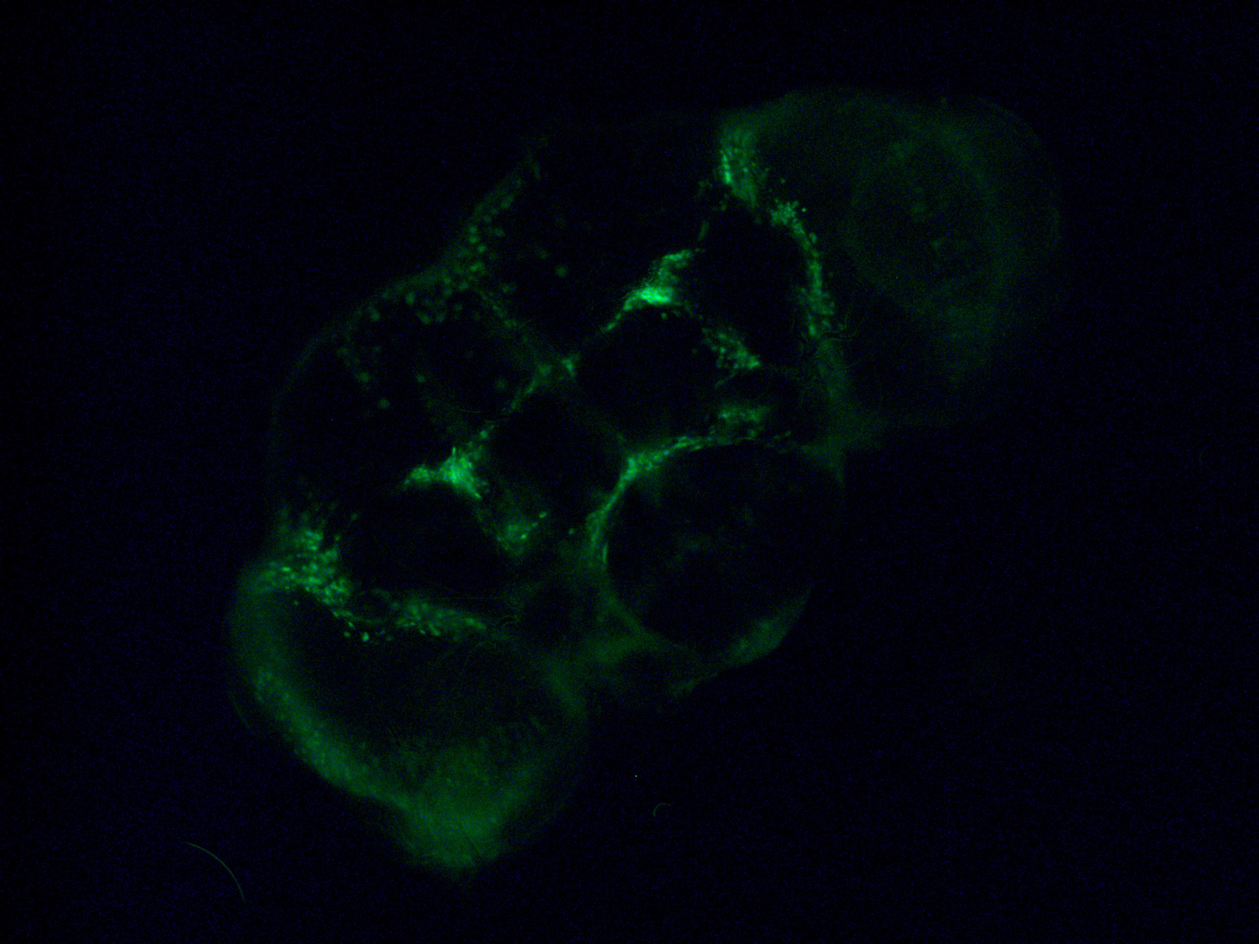
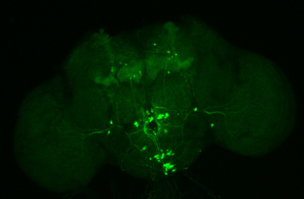
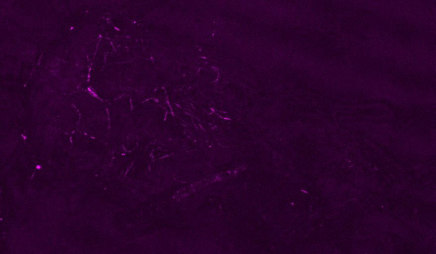
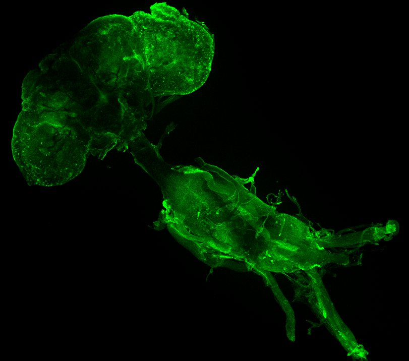
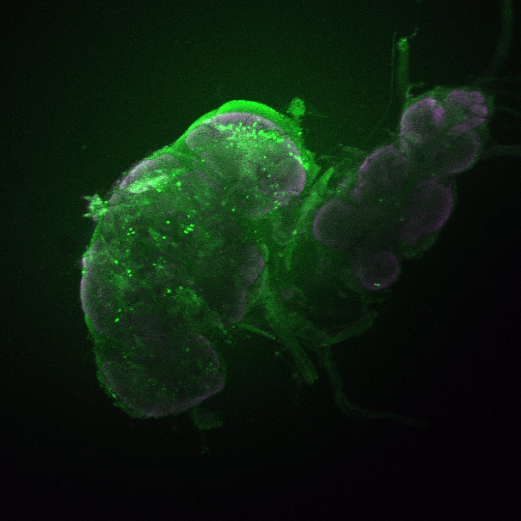
Axel stainning from c105-G4 alone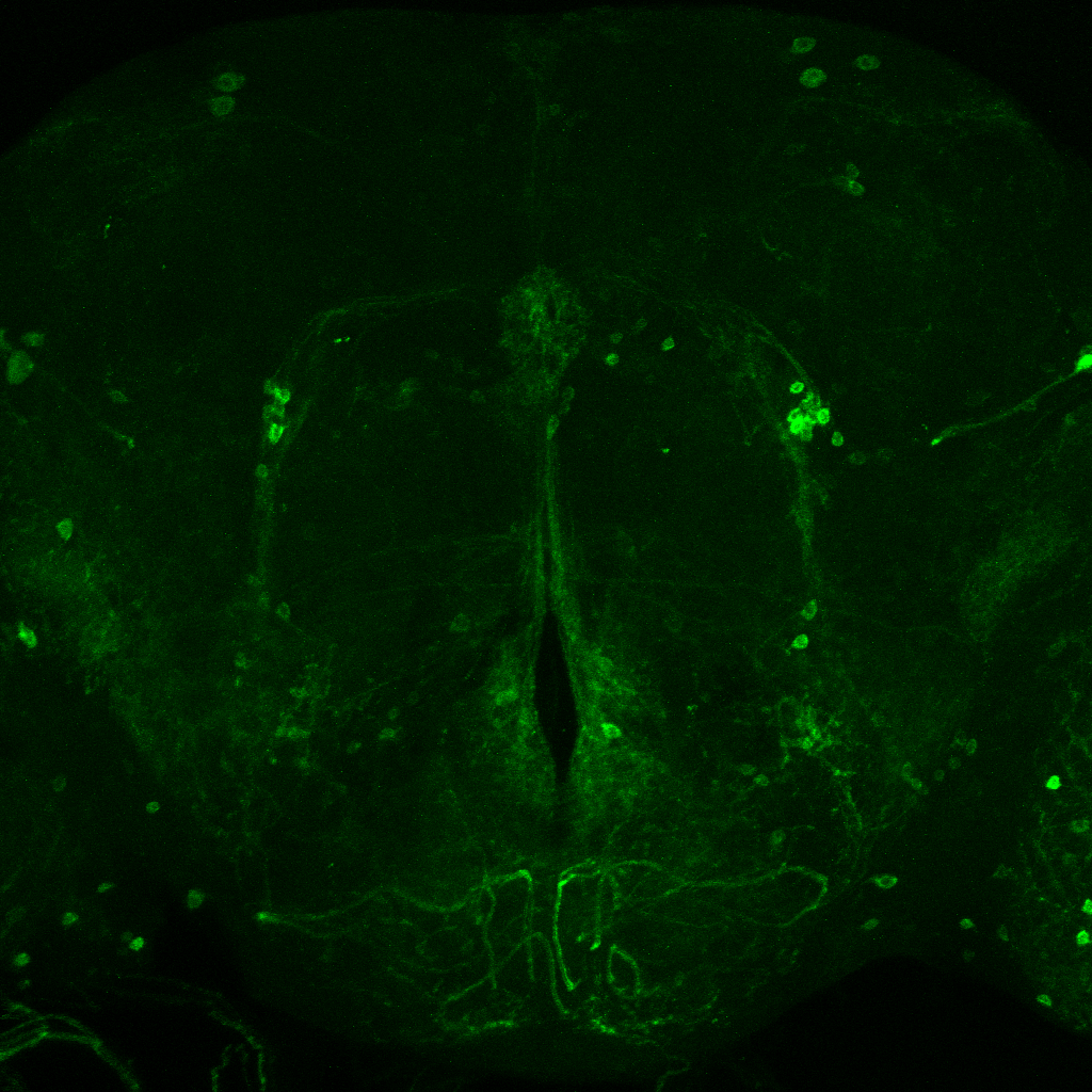
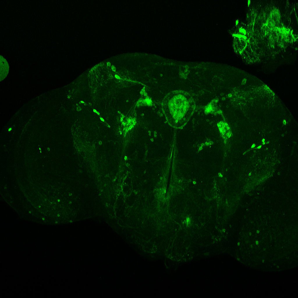
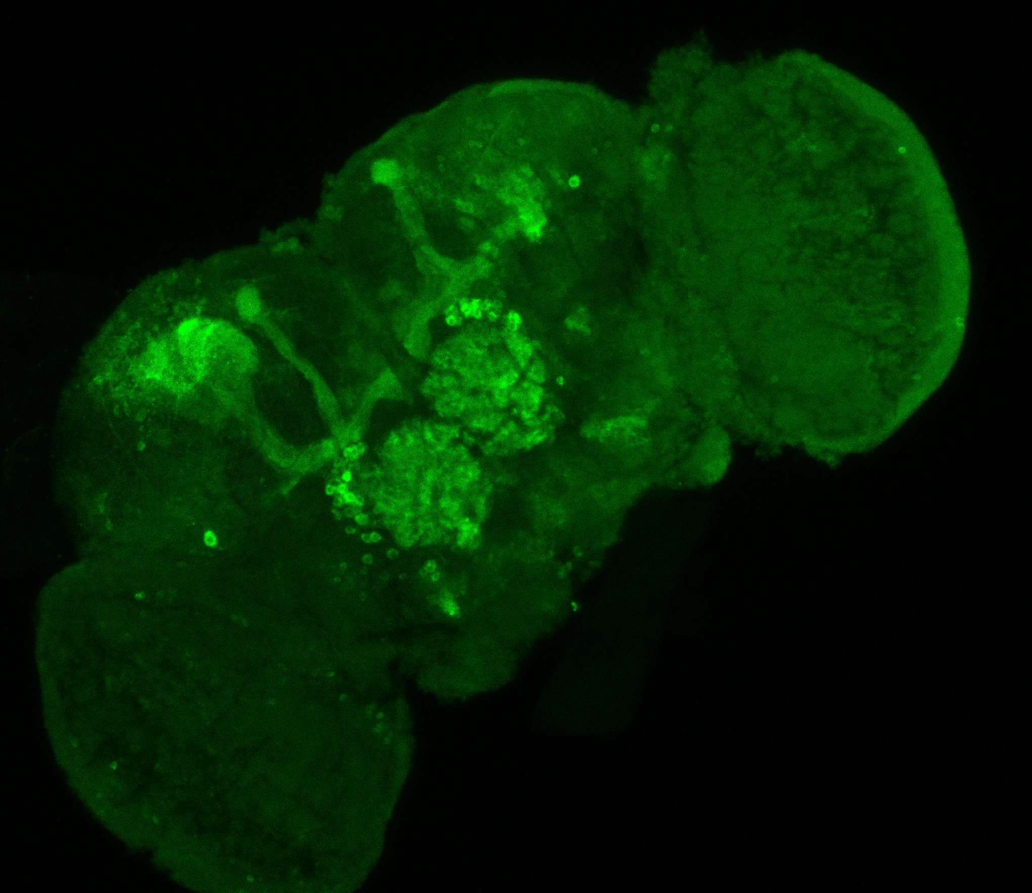
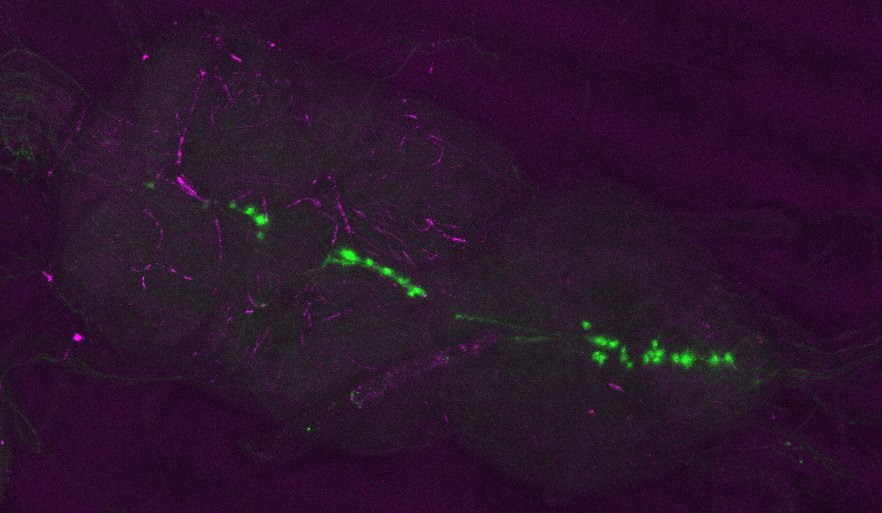
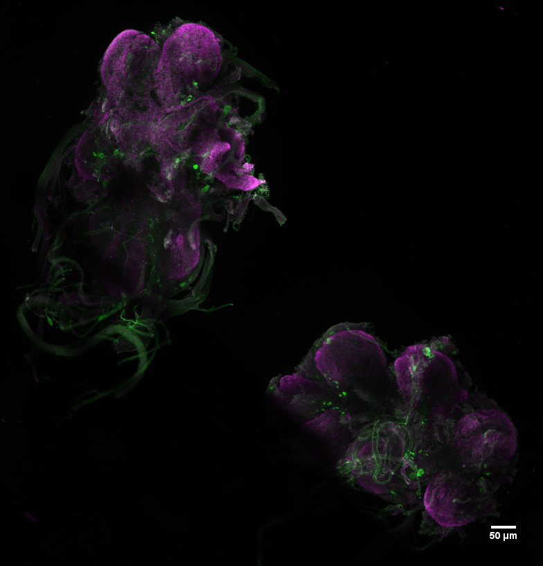
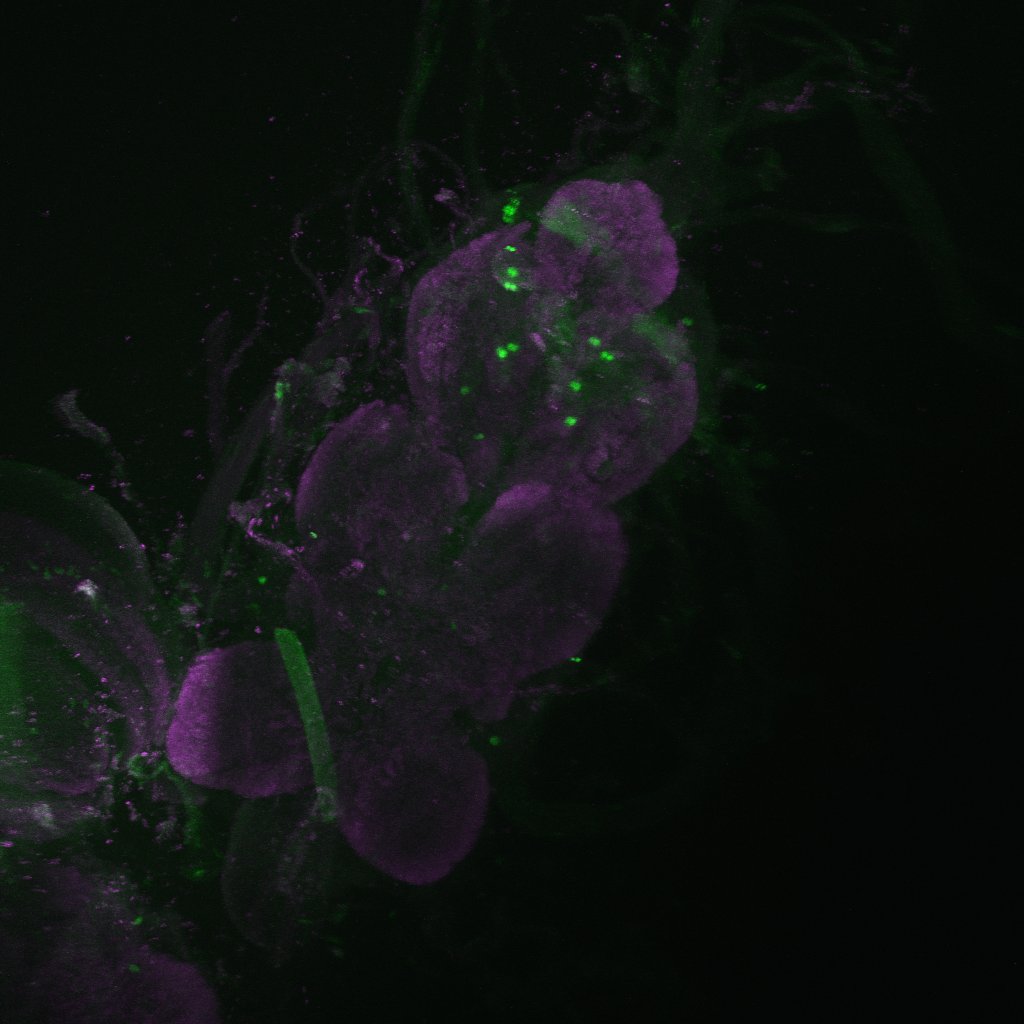
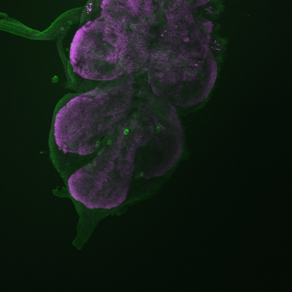
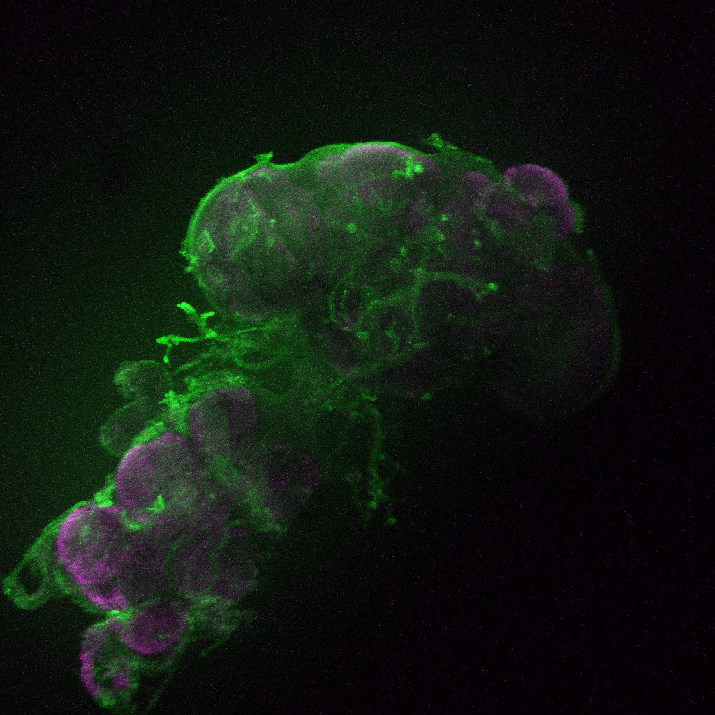
Axel stainning from both drivers together. I would say it really contains both driver lines.
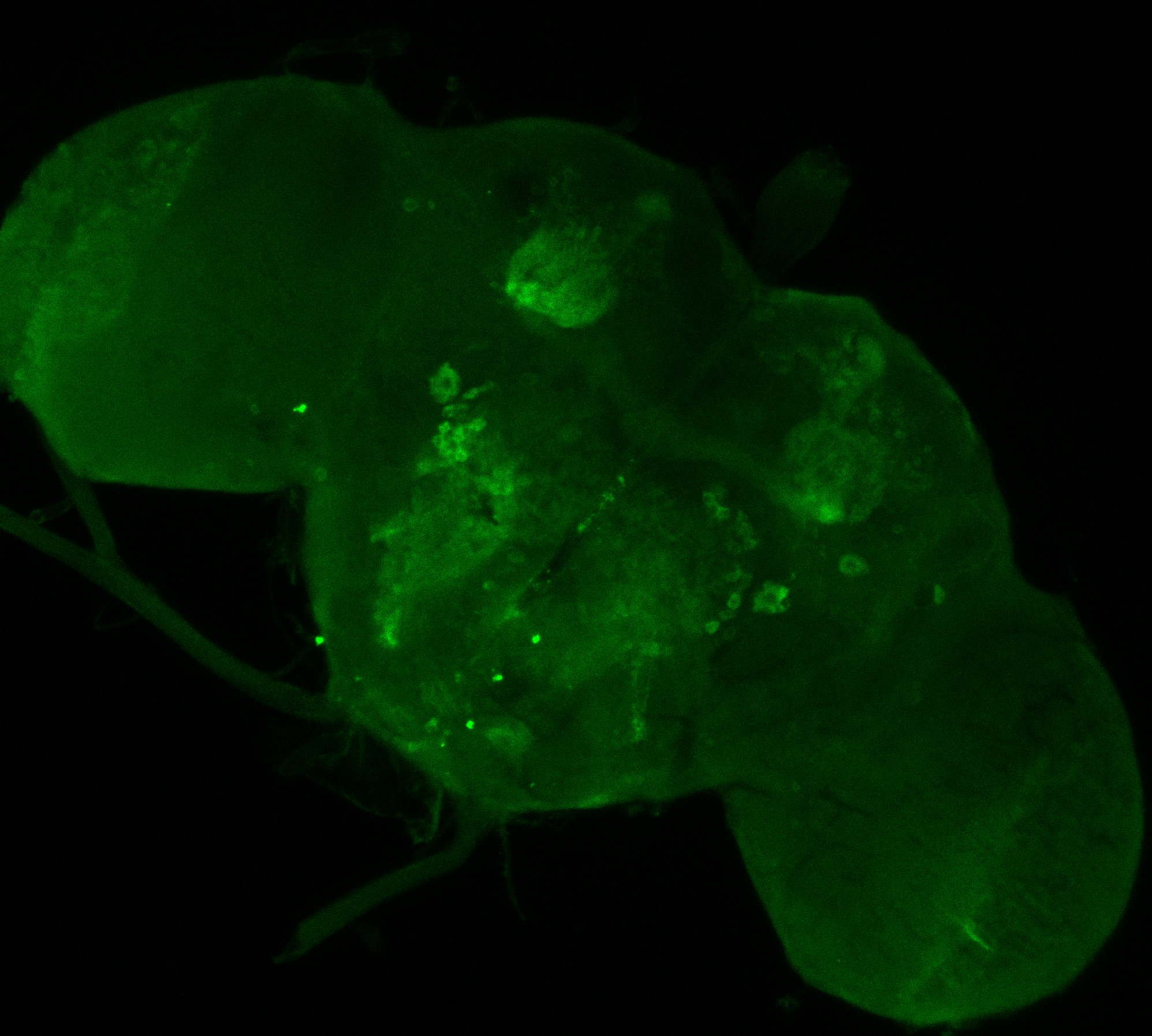
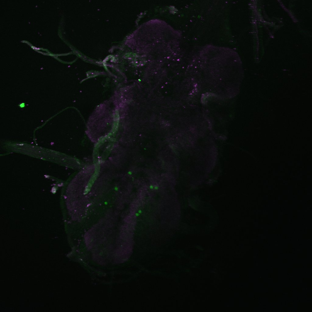
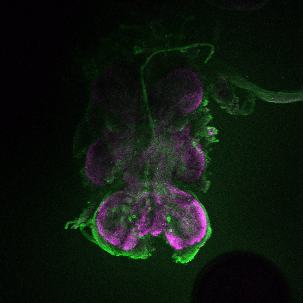
This are both driver lines together as well from Axel. To me it seems that only c105 is present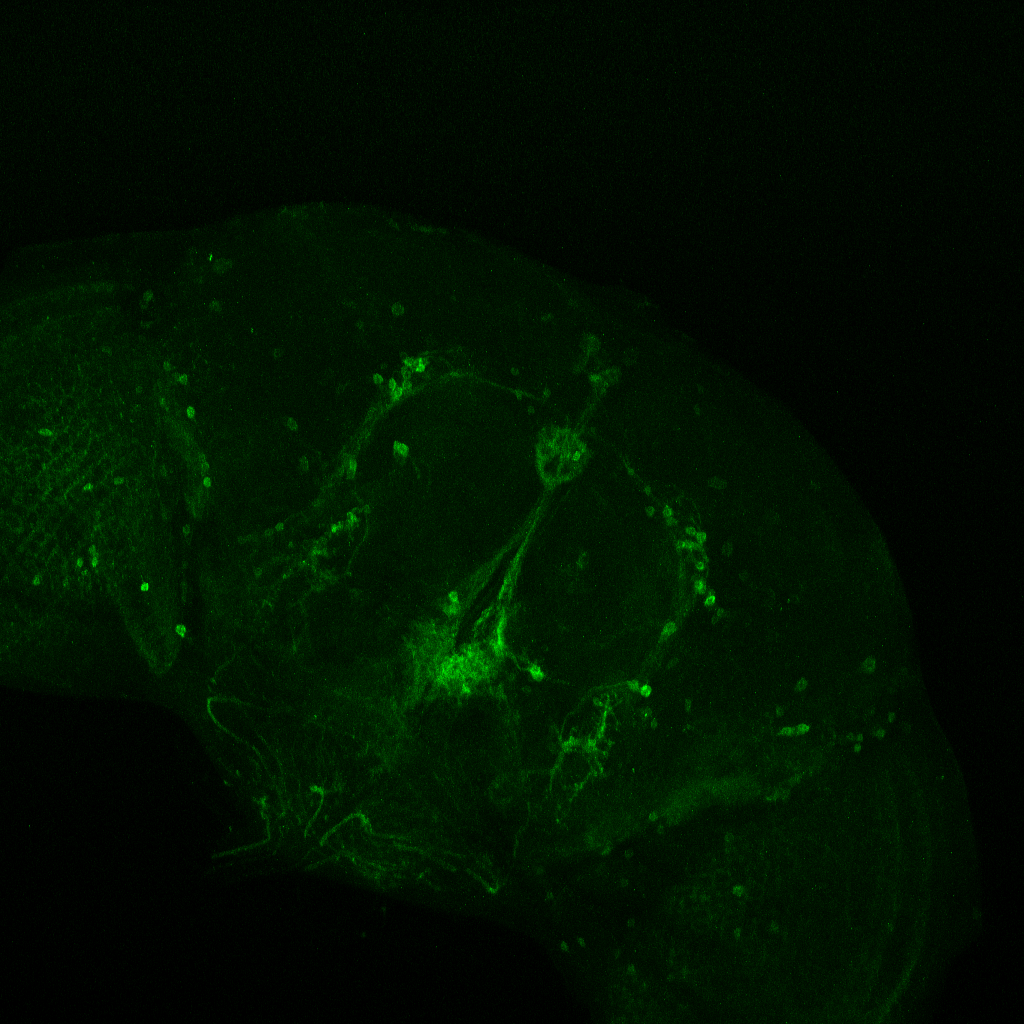
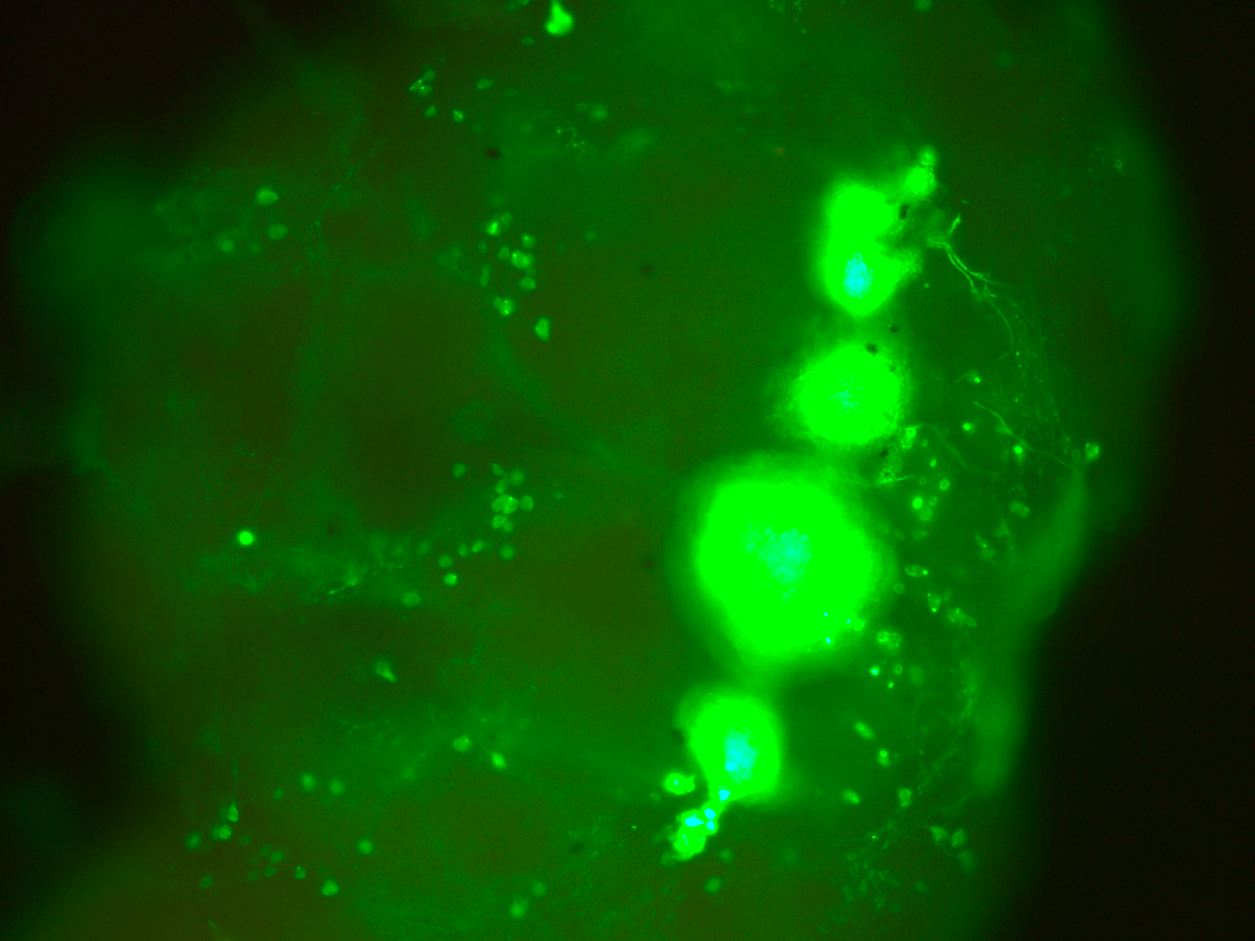
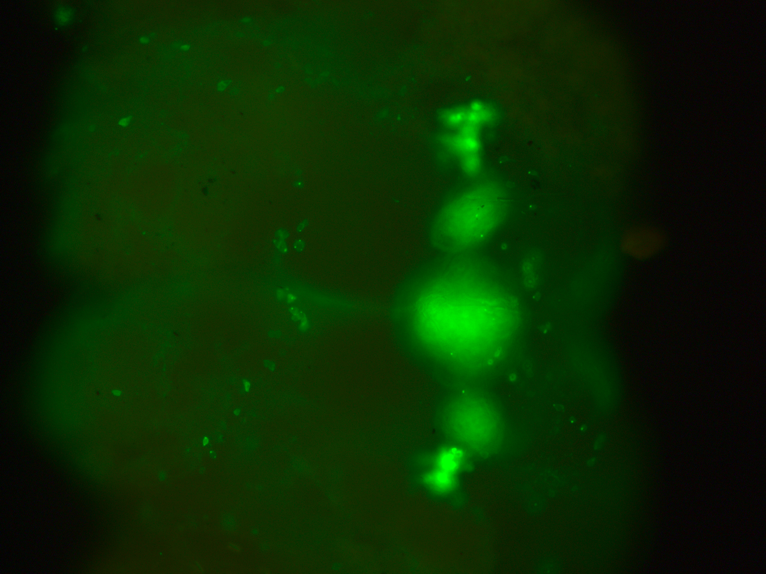
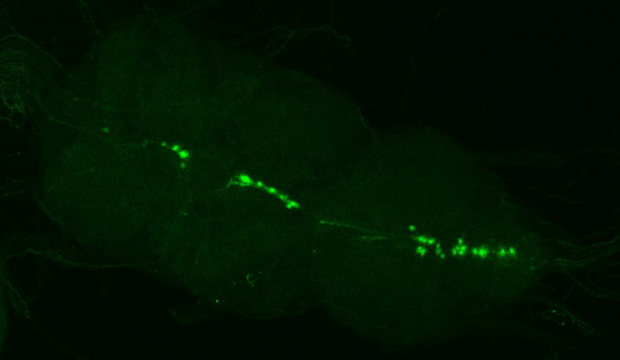
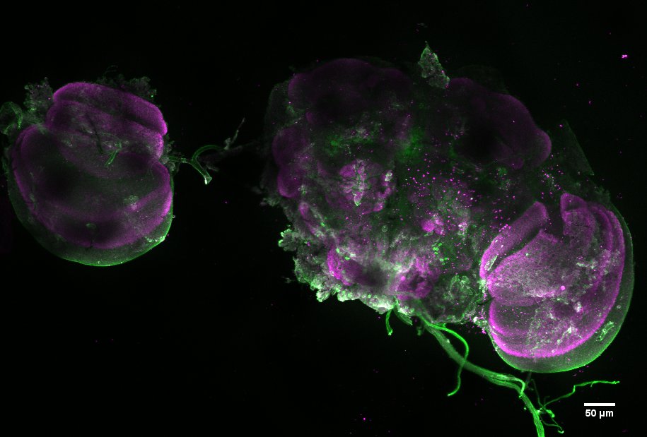
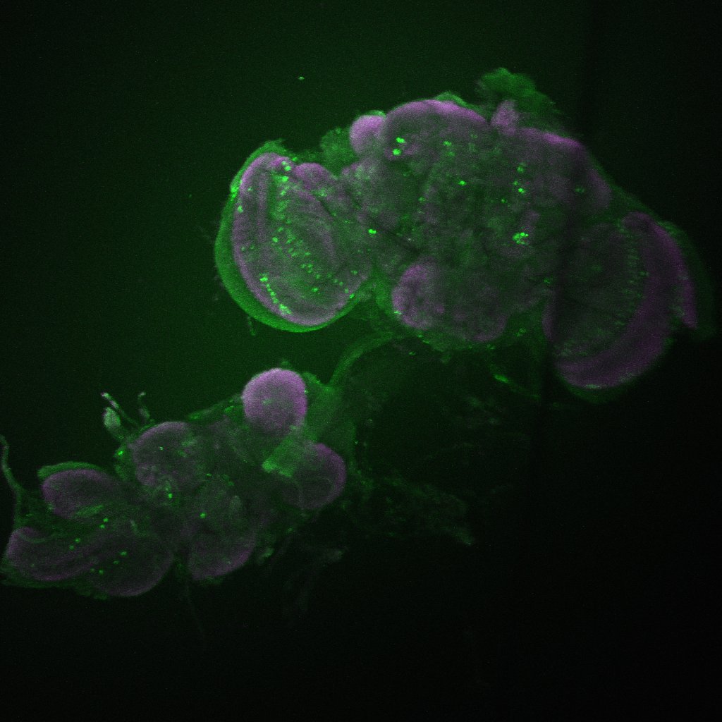
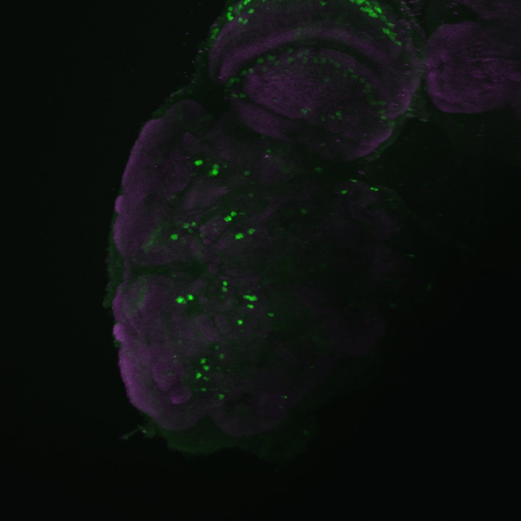
This are my stainnings at the fluorescence microscope (no confocal). This is to show that in all of the 10-12 brains I have looked at, they all had the c232 pattern present
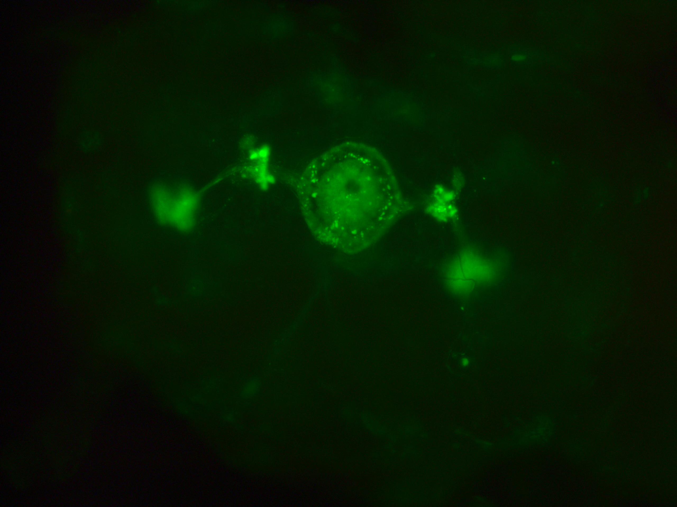
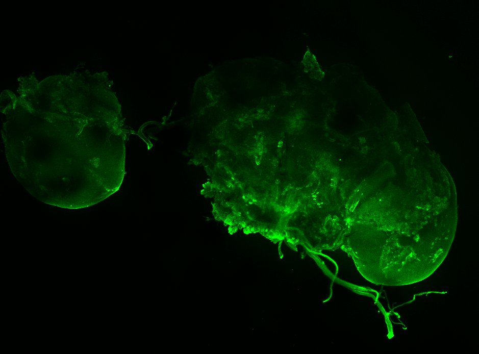
In addition, they had many more neurons outside from the central complex which I believe belong to the c105-G4 line. This is my only proof to show that c105 is also present, since the R1 neurons seem to be hidden when R2 neurons are stained.

I was also looking to the youtube video you have online, Björn. To me it seems I can only see the R1 ring neuron from the c105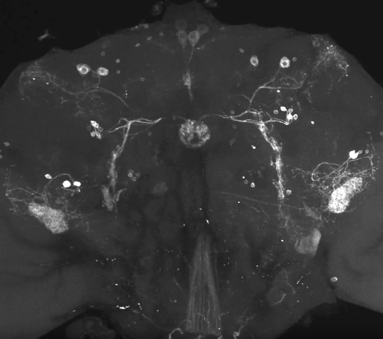

Category: Anatomy, flight, Spontaneous Behavior | No Comments
Testing CaLexA
on Monday, October 30th, 2017 1:32 | by Axel Gorostiza
Here I used the CaLexA tool with the elav driver, and reared the flies on constant darkness or under L-D cycles.
L-D Cycle
D-D
Category: Anatomy, wing clipping | No Comments
TbH MIMIC
on Monday, October 9th, 2017 2:28 | by Axel Gorostiza
Category: Anatomy, Biogenic Amines | No Comments
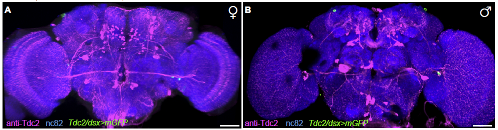
TbH-LexAs and TDC2-Gal4 comparison
on Monday, March 20th, 2017 2:17 | by Axel Gorostiza
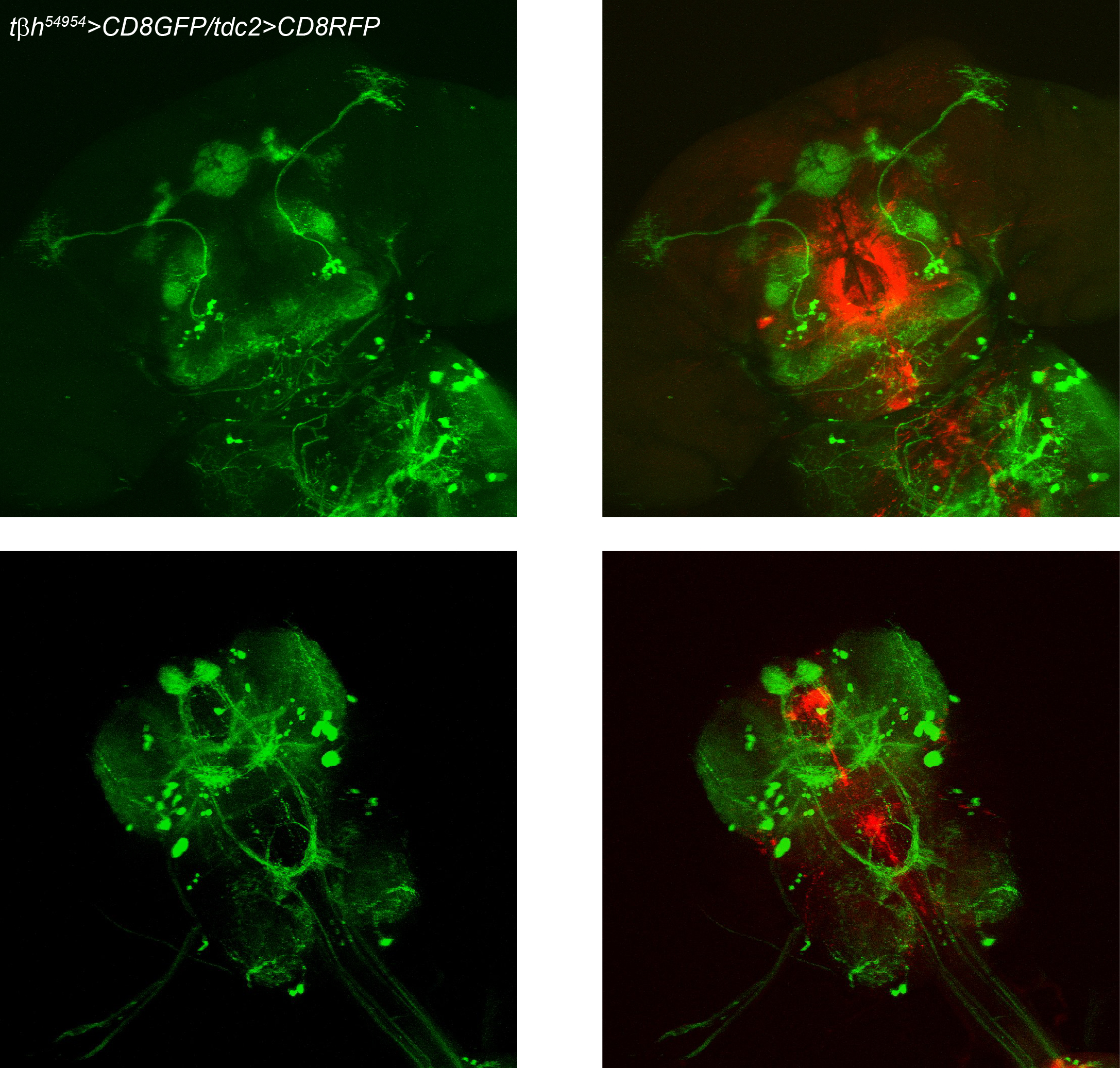
I recently combined the two TbH-lexA lines (54954 & 54075) with CD8GFP, and the TDC2-GAL4 line with CD8RFP, in order to compare their expression patterns. Here I present some of the dissections. The confocal is not working properly, but it is relatively good to draw some conclusions.
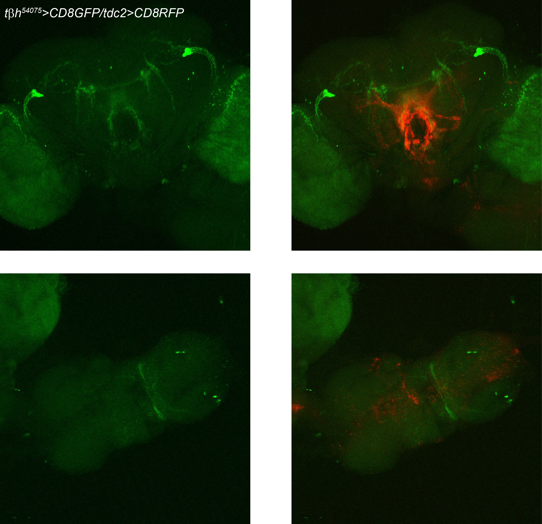
TDC2>GFP and anti-TβH (Scholz’s Lab)
TDC2>GFP (Gerber’s Lab)
anti-TDC2 (Goodwin’s Lab)
Category: Anatomy, Biogenic Amines, wing clipping | No Comments
DA neuronal populations and photopreference (counting neurons)
on Monday, March 13th, 2017 3:01 | by Axel Gorostiza
After refining my DA screening, I end up having three interesting GAL4s which lead to changes in photopreference after expressing Shibire and rising the temperature. What I am trying to do now is to understand if they label the same neuronal population or not.
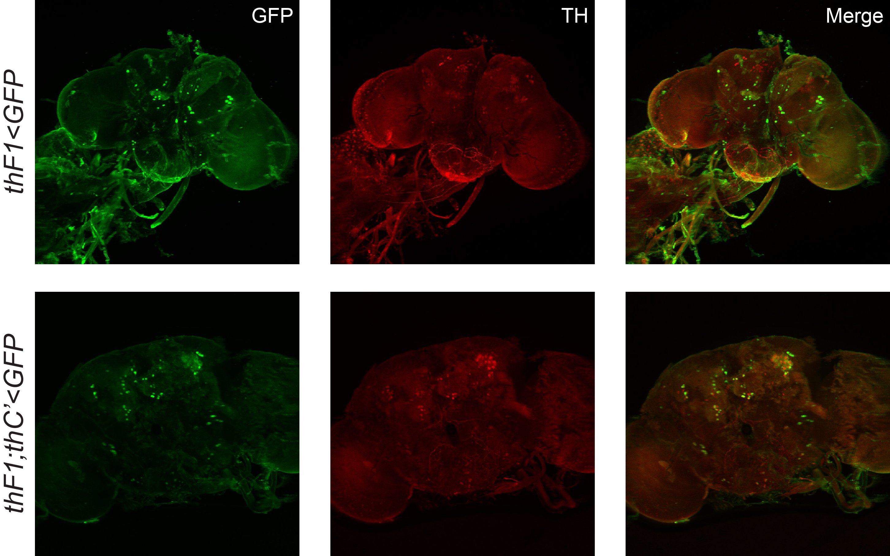
| Genotype | PAM | PAL | PPM1 | PPM2 | PPM3 | PPM4 | PPL1 | PPL2 | VUM |
| thF1>GFP | 0 | 0 | 0,25 | 3,75 | 4,25 | 0 | 3 | 0,75 | 0 |
| thF1;C’>GFP | 0 | 0 | 1 | 7 | 5 | 0 | 2,5 | 5,5 | 1,5 |
Category: Anatomy, Biogenic Amines, wing clipping | No Comments
TbH QF system
on Monday, September 12th, 2016 1:37 | by Axel Gorostiza
In order to study the role of OA and DA in photopreference I am constantly looking for new drivers that label subpopulations of these groups. I recently found a TbH driver from the QF system, and wanted to know how representative of the TDC2-G4 neurons was. I have established a QUAS-mCherry;TbH-QFs line (Magenta) and crossed it with TDC2-G4;UAS-GFP (Green). In both cases, what is shown is the endogenous expression.
Category: Anatomy, Biogenic Amines, wing clipping | No Comments
Octopaminergic neurons and phototactic flexibility
on Monday, February 1st, 2016 12:46 | by Axel Gorostiza
In previous experiments, I found one TβH(lexA)>shiTS combination that recapitulated TDC2>shiTS T-Maze results (https://lab.brembs.net/2015/09/looking-for-the-da-oa-neurons-involved-in-phototactic-flexibility/). Here I present the expression pattern of those two TβH-lexA drivers used.
TβH54954-LexA (Brain)
TβH54954-LexA (VNC)
TβH54075-LexA (Brain & VNC)

Category: Anatomy, Biogenic Amines, wing clipping | No Comments
TH-F1 anatomy
on Monday, December 7th, 2015 2:22 | by Axel Gorostiza
I am starting to study in more detail the genotypes that were positive in the DA screen. One was the TH-F1. Here I show the anatomy. I had several problems with the old confocal. I will try to use only the new one.
Category: Anatomy, wing clipping | No Comments
Phototactic flexibility – Neural substrates
on Monday, November 23rd, 2015 2:29 | by Axel Gorostiza
In order to find which dopaminergic and octopaminergic neurons are related to light preference and the switch on it seen after clipping the wings, I decided to use a tool called CaLexA.
Here we can see my first attempt to use it. We can see the CNS from TH>CaLexA flies with and without wings.
I did not see any special signal in the brains, but I still have to play around a little bit more.
With Wings
Without Wings
Category: Anatomy, Biogenic Amines, wing clipping | No Comments
“Virtual brain” site
on Saturday, March 2nd, 2013 4:00 | by Christine Damrau
Arnim Jenett (Janelia Farm Research Campus), Kazunori Shinomiya, Kei Ito (both Tokyo University), and other anatomists made a great site with a 3D-viewer of adult Drosophila brains available. You have the chance to scroll threw a whole mount stack while ticking different brain areas. Those brain areas are listed next to the stack. Different areas are coloured differently, so that you can look at the location of several areas in the same brain. On the main page you can find simply explained tutorials about the usage of the site. It is correlated to the anatomical search engine of the Janelia farm GAL4 collection.
Because it was very helpful to me to learn all the synonyms of relevant areas and because I think it is very helpful to learn more about the structure of the Drosophila brain in general I wanted to advertise the site here.
https://www.virtualflybrain.org/site/vfb_site/overview.htm
Category: Anatomy, open science | 1 Comment