aPKC/FoxP colocalization confocal images
on Friday, January 23rd, 2026 12:52 | by Fridrik Kjartansson
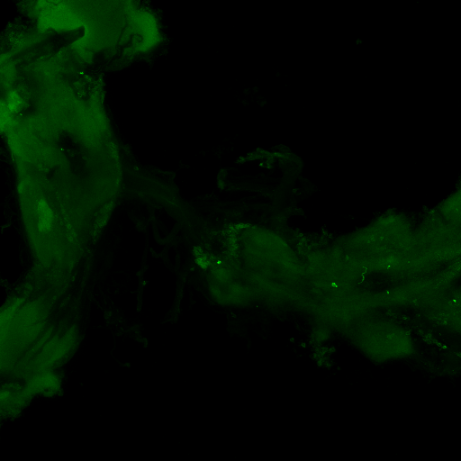

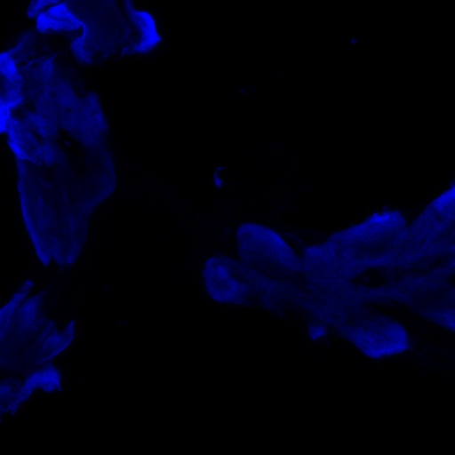
Staining is a bit faint and brain quality a bit sub-optimal and no punctae can be observed in the FoxP channel, decreasing the gain did not resolve this, blocking was done for 1 hour at RT. Perhaps increasing concentration of 1st degree antibodies, especially for RFP might improve the results.

Category: Anatomy, crosses, Foxp, PKC, PKC_localisation
Leave a Reply