Cross-reactive anti-HSP70 binding and secondary antibody effects on the protein marker
on Monday, October 5th, 2026 1:20 | by Jana Handfest


Category: HSP70 | No Comments
HSP70 expression after longer light-induced activation of chrimson
on Sunday, October 4th, 2026 3:28 | by Annika Schmid

Expression of HSP70 (70 kDa, detected using fluorescence at 700 nm (red)) after:
- 1h of heat shock at 37 °C (WTB, positive control)
- 15, 30 and 60 min of red-light exposure (after 2 days of feeding with ATR)
- various negative controls (ethanol/ water instead of ATR for 60 min/ 0 min, and ATR for 0 min)
HSP70 expression levels were normalized to the housekeeping protein actin (42 kDa, detected by fluorescence at 800 nm (green)).

Category: HSP70, Uncategorized | No Comments
Quantification of HSP70 increase after heatshock
on Sunday, October 4th, 2026 2:41 | by Annika Schmid


Male and female groups after heat-shock (HS) and non-stress (n.S.) conditions. HSP70 was detected using a rat anti-HSP70 primary antibody and a donkey anti-rat secondary antibody with fluorescence detection at 700 nm (red). Actin was used as loading control (intensity slightly edited) and detected using a rabbit anti-actin primary antibody and a goat anti-rabbit secondary antibody at 800 nm (green). HSP70 signal intensity was quantified and normalized to the corresponding actin signal.

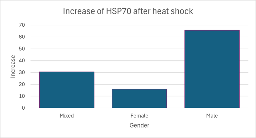
Increase of HSP70 expression after heatshock. Difference in increase between sexes is not significant (t-test, p-value = 0.3).
Category: HSP70 | No Comments
HSP70 expression after light-induced activation of chrimson
on Tuesday, September 29th, 2026 7:47 | by Annika Schmid
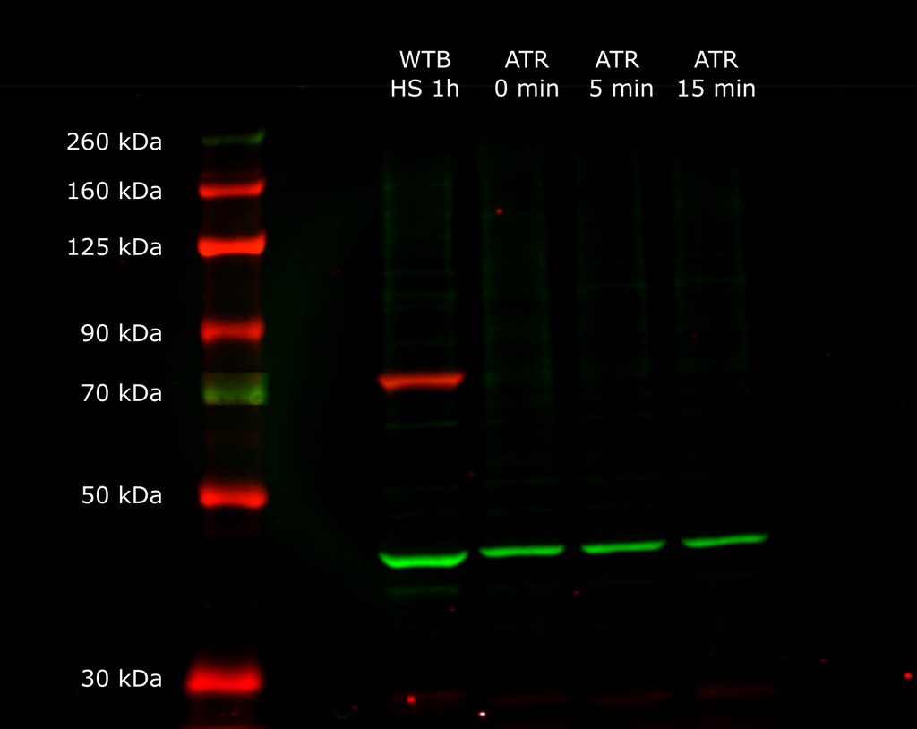
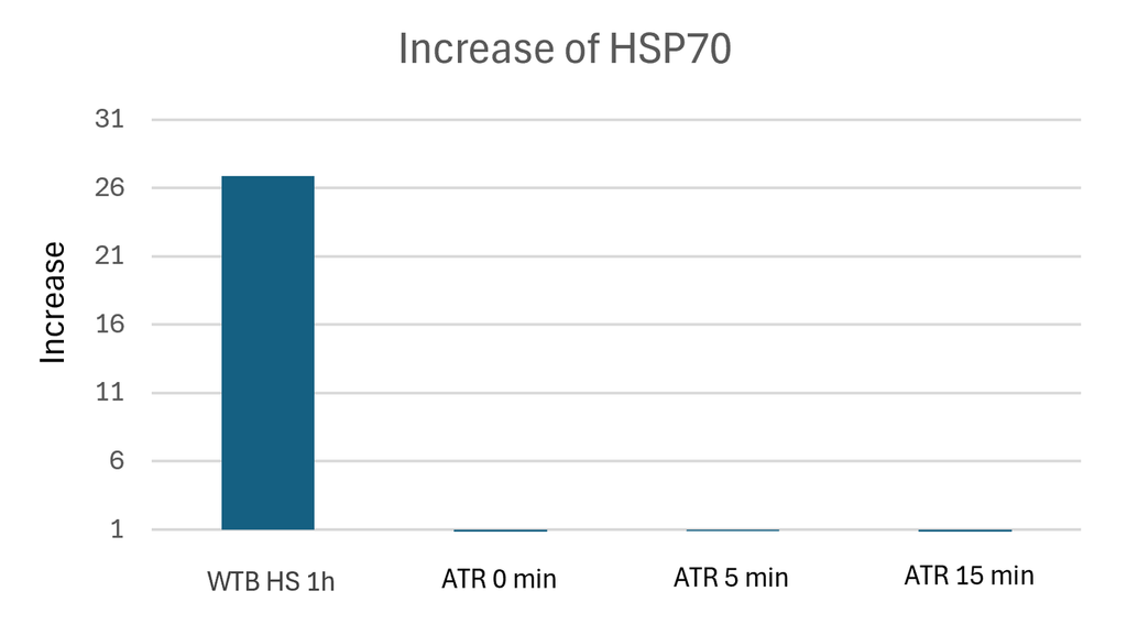
Expression of HSP70 (70 kDa, detected using fluorescence at 700 nm (red)) after:
- 1h of heat shock at 37 °C (WTB, positive control)
- 0 min of red-light (660 nm) exposure (negative control)
- 5 min of red-light exposure
- 15 min of red-light exposure
HSP70 expression levels were normalized to the housekeeping protein actin (42 kDa, detected by fluorescence at 800 nm (green)).
Category: HSP70 | No Comments
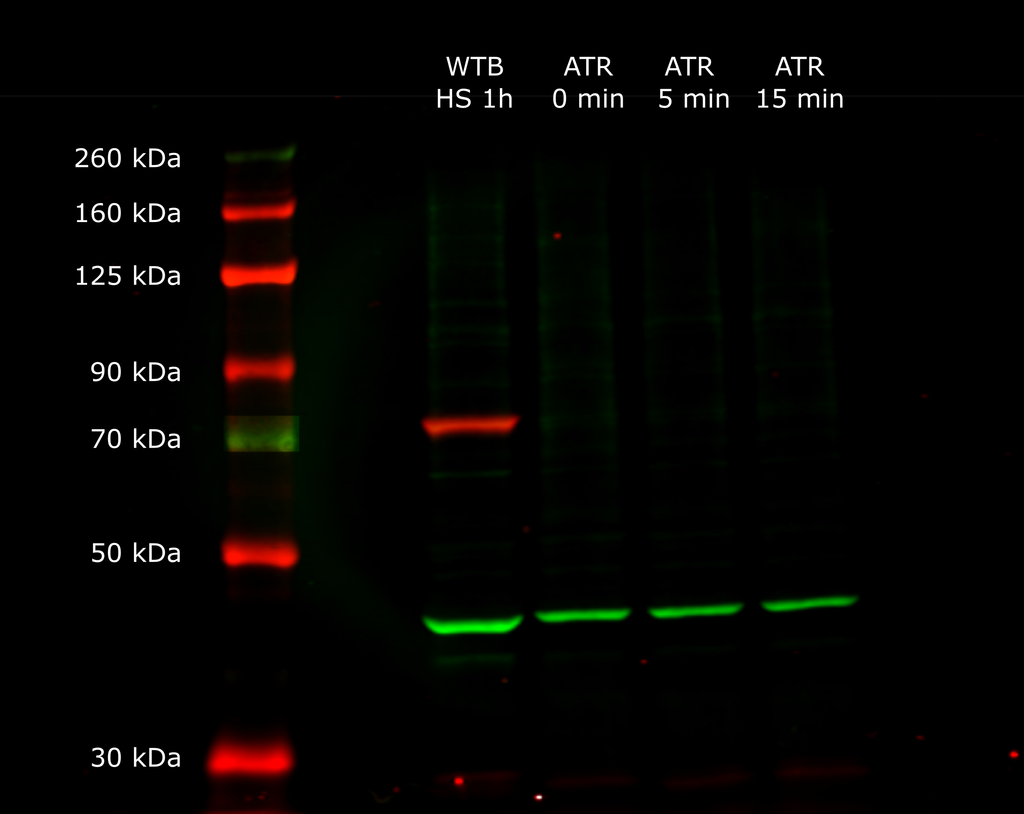
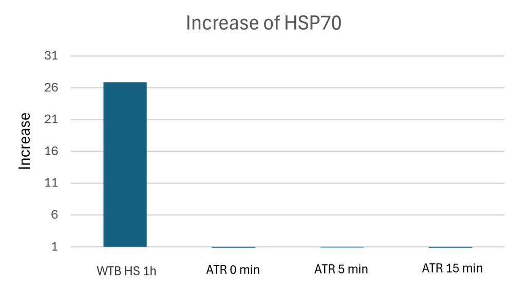
Light-induced activation of Chrimson does not induce HSP70 expression
on Monday, September 28th, 2026 1:16 | by Jana Handfest
Category: HSP70, Uncategorized | No Comments
Western blot analysis and quantification of HSP70 expression in response to heat shock
on Wednesday, September 23rd, 2026 10:35 | by Annika Schmid
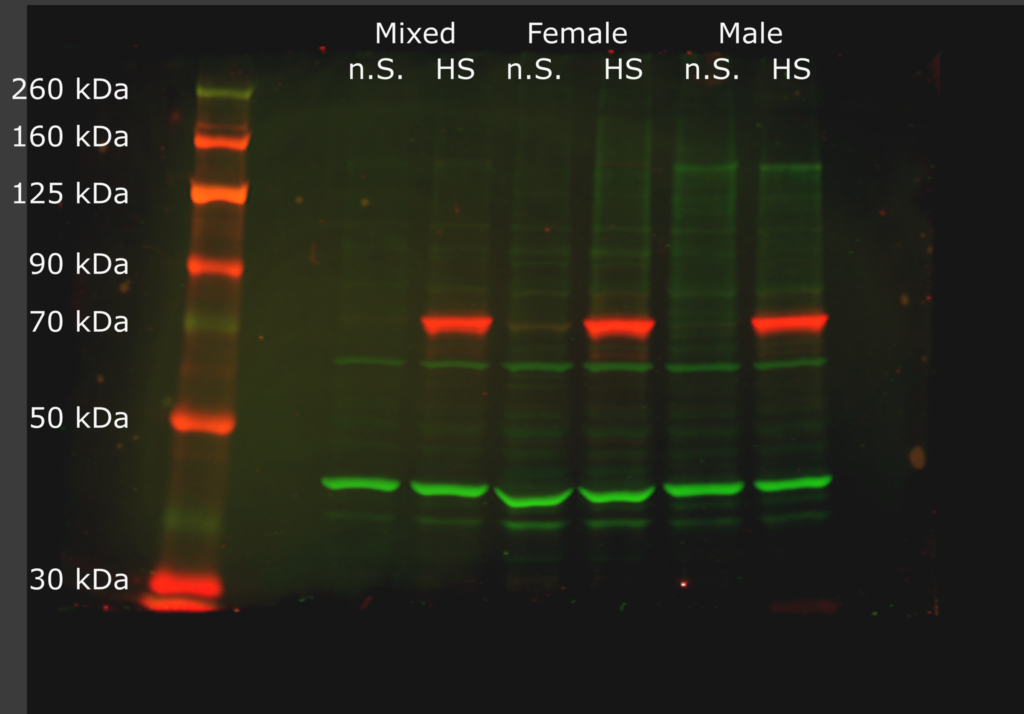
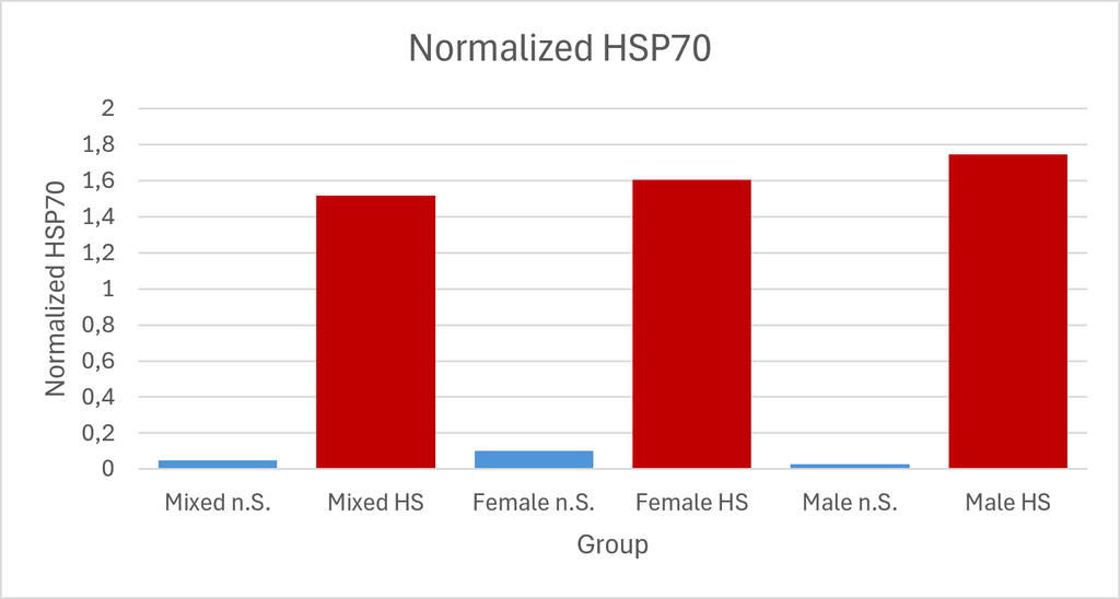
Male, female, and mixed groups under heat-shock (HS) and no-stress (n.S.) conditions. HSP70 was detected using a rat anti-HSP70 primary antibody and a donkey anti-rat secondary antibody with fluorescence detection at 700 nm (red). Actin was used as loading control and detected using a rabbit anti-actin primary antibody and a goat anti-rabbit secondary antibody at 800 nm (green). HSP70 signal intensity was quantified and normalized to the corresponding actin signal.
Category: HSP70 | No Comments
Western blot analysis and quantification of HSP70 expression in response to heat shock
on Monday, September 21st, 2026 12:53 | by Jana Handfest
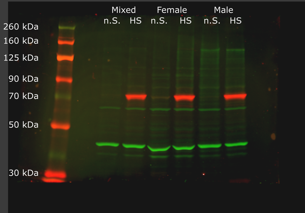
Six experimental groups were analyzed, consisting of male, female, and mixed samples under heat-shock (HS) and no-stress (n.S.) conditions. For heat-shock-treatment, samples were exposed to 37 °C for 1 h, followed by a 1 h recovery period at 25 °C. Control samples were treated exactly the same but not exposed to heat stress.
Western blot analysis was performed to determine HSP70 protein levels. HSP70 was detected using a rat anti-HSP70 (1:3000) primary antibody and a donkey anti-rat (1:10000) secondary antibody with fluorescence detection at 700 nm (red). Actin was used as loading control and detected using a rabbit anti-actin (1:8000) primary antibody and a goat anti-rabbit (1:10000) secondary antibody at 800 nm (green). HSP70 signal intensity was quantified and normalized to the corresponding actin signal.
A strong HSP70 signal was observed in all heat-shock groups, indicating a clear induction of HSP70 in all heat shocked groups and only very low levels of HSP70 in the no stress groups.
The male and female groups showed different increases of HSP70 expression following heat shock, with higher levels observed in males. This result was unexpected, as previous literature suggests that males may constituively express HSP70 in spermatogonia during certain development stages, which could have resulted in higher basal HSP70 signal in the male samples. In contrast, females are generally considered to have an higher heat tolerance, which may contribute to the comparatively smaller increase in HSP70 expression between the no stress and heat shock conditions detected in females. Interestingly, however, basal HSP70 levels in female no stress group were higher than those observed in males.
After the experiment, we noticed, that there were differences in the number of flies per tube used during the heat shock and recovery periods. Therefore, some groups may have experienced additional stress due to overcrowding, which could have incluenced HSP70 expression. Consequently, the observed differences cannot yet be attributed exclusively to the gender. To determine whether the differences are genuinely gender-dependent, the experiment will be repeated under standardized conditions.
Category: HSP70 | No Comments
HSP70 after heat shock in western blot
on Monday, September 14th, 2026 12:50 | by Annika Schmid
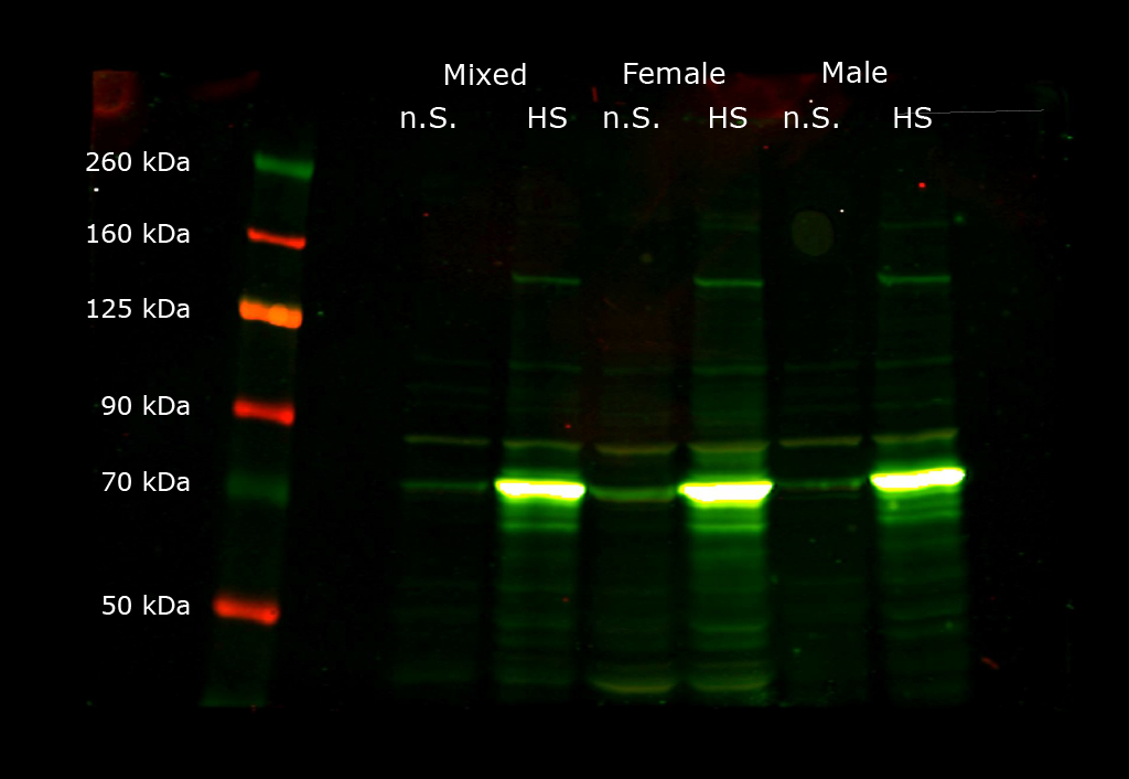
Expression of HSP70 (70 kDa) after heatshock (HS) or no heatshock (n.S.) in groups of only female, male and both genders (mixed). Fluorescence was measured at a wavelength of 700 nm (anti-mouse) and 800 nm (anti-rat).
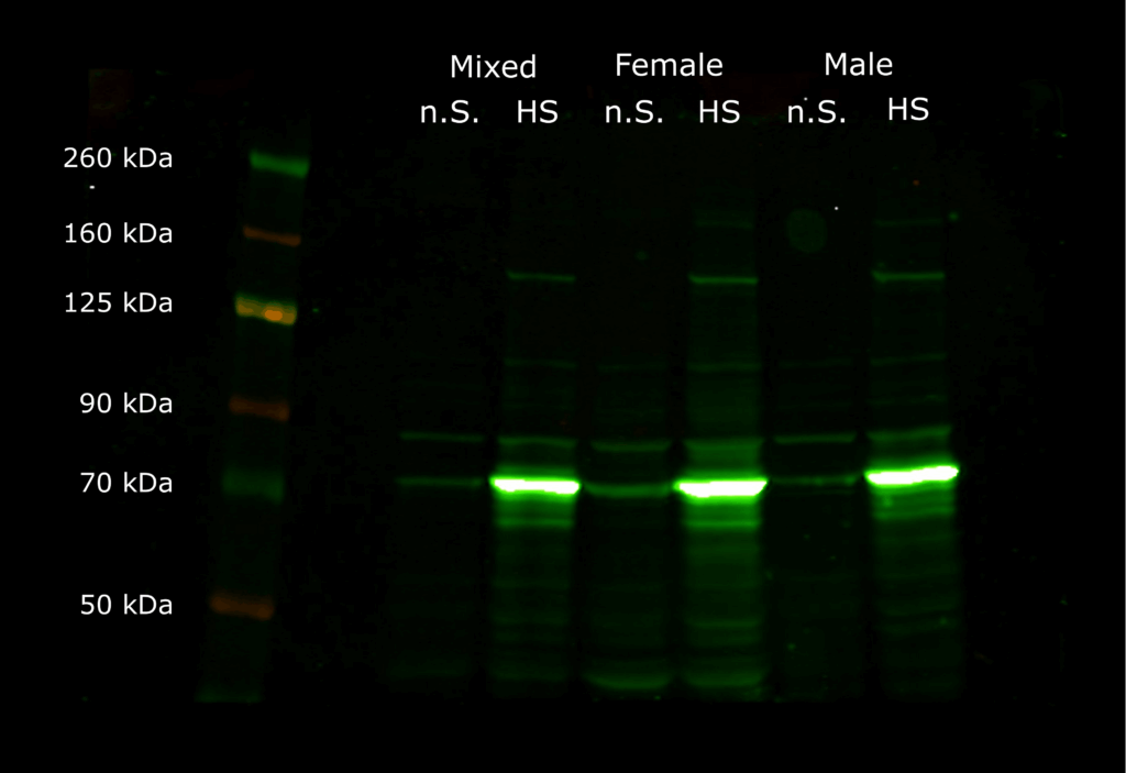
Expression of HSP70 (70 kDa) after heatshock (HS) or no heatshock (n.S.) in groups of only female, male and both genders (mixed). Fluorescence was measured at a wavelength of 800 nm (anti-rat).
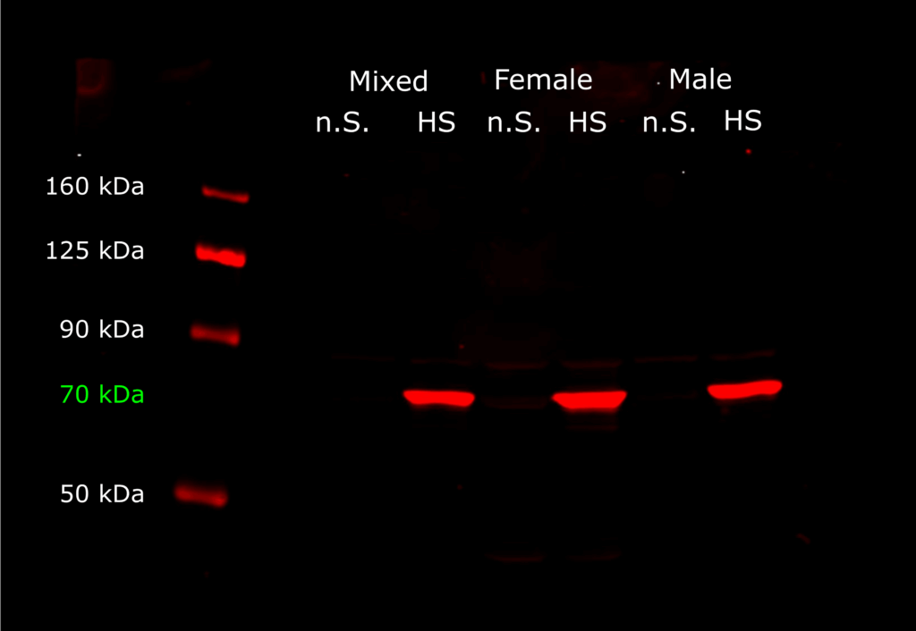
Expression of HSP70 (70 kDa) after heatshock (HS) or no heatshock (n.S.) in groups of only female, male and both genders (mixed). Fluorescence was measured at a wavelength of 700 nm (anti-mouse).
Category: HSP70 | No Comments